Learning Objectives
By the end of this chapter you should be able to:
- Define early, mid and late rehabilitation using tissue biology and function rather than calendar time.
- Identify objective criteria that justify progression between phases.
- Apply dosage parameters and pain-monitoring rules at each stage.
- Recognise red flags and contraindications that stop progression.
Why a Continuum, Not a Timetable
Rehabilitation is a continuous process of matching load to tissue capacity. Phases are labels for points along that continuum. Time since injury or surgery sets only the earliest possible date for progression. Meeting criteria sets the actual date. Athletes who progress by criteria rather than time reinjure less often (Grindem et al., 2016; Kyritsis et al., 2016).
Pathophysiology: Healing Biology and the Envelope of Function
Soft-tissue repair overlaps across three biological stages:
| Stage | Approx. timeframe | Key events | Clinical implication |
|---|---|---|---|
| Inflammatory | 0–7 days | Haemostasis, neutrophil then macrophage infiltration | Protect, limit effusion, maintain unaffected tissue |
| Proliferative | ~3 days–6 weeks | Fibroblast activity, type III collagen, angiogenesis | Graded, controlled loading to direct fibre alignment |
| Remodelling | ~3 weeks–12+ months | Type III replaced by type I collagen, with cross-linking and realignment | Progressive heavy and high-velocity loading |
Tendon, ligament and grafted tissue remodel slowly. Strength and function can recover faster than structural maturity, which is why athletes often feel ready before their tissue is. Dye's envelope of function describes the range of load a tissue tolerates without breakdown. Injury narrows the envelope. Rehabilitation aims to widen it progressively without exceeding it. Under-loading causes deconditioning, and over-loading causes setbacks.
Neuromuscular changes run alongside tissue healing. These include arthrogenic muscle inhibition (especially quadriceps after knee injury), altered motor control, and fear-related movement avoidance.
Defining the Phases
Early phase (protect and restore baseline)
- Goals: control pain and effusion, restore range of motion (ROM), activate inhibited muscle, preserve fitness, and normalise gait.
- Typical features: pain at rest or with ADLs, visible effusion, antalgic gait, marked strength deficit (limb symmetry index, LSI, <60%).
Mid phase (rebuild capacity)
- Goals: restore full ROM, build strength and muscle mass, restore single-leg control, and begin low-level impact and running.
- Typical features: no effusion at rest, pain ≤3/10 with loading, strength LSI 60–85%.
Late phase (reconditioning to sport demands)
- Goals: restore maximal strength and power, reactive and sport-specific agility, high-speed running, and psychological readiness, leading to return to training and then return to competition.
- Typical features: LSI approaching 90%, with residual deficits in rate of force development, fatigue resistance and confidence.
The Transition Window
Early-to-mid and mid-to-late transitions are the points where most mistakes occur. Athletes feel good and function well in daily life, but the tissue has not yet been challenged at sport intensity. The transition window opens when the criteria below are met. Progression then occurs through a short, overlapping period of graded exposure, usually 1–3 weeks, rather than abruptly.
Presentation: Recognising Where the Athlete Sits
Look for these at the transition points:
- Ready to leave early phase: no more than trace effusion, pain ≤3/10 during ADLs, near-normal gait, and ROM within 5–10° of the other side.
- Ready to leave mid phase: no pain or swelling response 24 hours after the heaviest session, and competent single-leg tasks.
- Not ready: a delayed pain or swelling flare (next morning), compensatory trunk lean, dynamic knee valgus, or reluctance to land on the injured limb.
Assessment: Tests, Measures and Normative Values
Combine impairment measures, functional tests and psychological readiness. No single test is sufficient.
Pain and irritability
- Numeric Pain Rating Scale (NPRS) during and 24 hours after loading.
- Palpation and the specific provocation tests for the injured structure.
Swelling
- Knee: stroke test (0 = none, trace, 1+, 2+, 3+). Aim for 0–trace before progression.
- Ankle: figure-of-eight girth. A difference of <1 cm is the usual target.
ROM
- Goniometry, with knee extension equal to the other side (0°, or hyperextension if that is the athlete's norm).
- Weight-bearing lunge test for the ankle. A common benchmark is ≥10 cm to the wall (about 35–40°), with <1.5 cm side-to-side difference.
Strength
- Handheld dynamometry or isokinetic testing at 60°/s and 180°/s.
- Target LSI ≥90%. Hamstring-to-quadriceps ratio is conventionally ≥60% at 60°/s.
- Hamstring injury: Nordic hamstring or isometric 90/90 testing, aiming for ≤10% asymmetry.
Functional and dynamic control
- Hop battery (single, triple, crossover, 6-m timed hop), each with LSI ≥90%.
- Y-Balance Test anterior reach asymmetry <4 cm.
- Single-leg squat, drop-jump landing assessment (look for knee valgus, trunk sway).
Psychological readiness
- ACL-RSI (a score ≥65 is a common threshold) or the Tampa Scale of Kinesiophobia.
Mid-to-late criteria summary
| Domain | Threshold |
|---|---|
| Effusion | 0–trace |
| Pain with sport-like loading | ≤2/10 and settled within 24 h |
| Quad/hamstring strength LSI | ≥80–85% (late phase target ≥90%) |
| Hop tests LSI | ≥80–85% (late phase target ≥90%) |
| ACL-RSI | ≥65 |
Management: Staged Protocols and Dosage
Early phase
- Frequency: daily, with 2–3 sessions per day of short, low-load exercises.
- Isometrics: 5 × 45 s at ~70% MVC for pain modulation (tendinopathy), or 5 × 10 s holds for muscle activation.
- ROM: 3 × 10–15 repetitions, up to the point of mild stretch.
- Neuromuscular electrical stimulation if there is quadriceps inhibition: 2–3 sessions per week at 50–75 Hz.
- Cardiovascular maintenance: upper body or non-weight-bearing cycling, 20–30 minutes at moderate intensity.
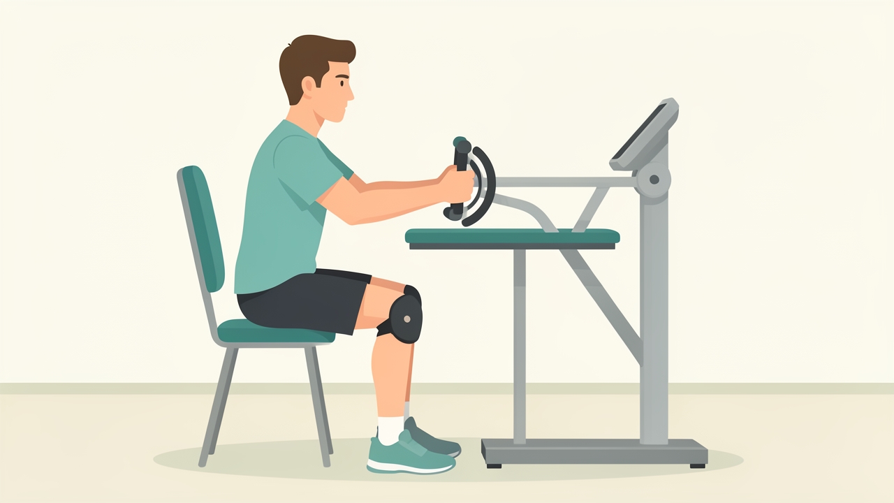
Early phase: preserve fitness with upper-body or non-weight-bearing cycling for 20–30 minutes at moderate intensity.
Mid phase
- Strength: 3–4 sets × 6–12 repetitions at 65–80% 1RM, 2–3 sessions per week, with 48 hours between sessions loading the same tissue.
- Tendon: heavy slow resistance, 3–4 sets progressing from 15RM to 6RM across ~12 weeks, 3 sessions per week.
- Impact: begin with 40–60 foot contacts per session, progressing to ~100.
- Running: begin only if walking is pain-free, effusion is absent, and the athlete can perform 20 single-leg heel raises and 10 single-leg squats to 60° with control. Use walk-jog intervals, for example 1:1 minutes for 20 minutes, 3 sessions per week on alternate days.
Late phase
- Strength and power: 3–5 sets × 3–6 repetitions at 80–90% 1RM, plus plyometrics (100–150 contacts per session, 2 sessions per week). Include rate-of-force-development work.
- Speed: progress running from 70% to 100% maximum speed over several sessions, with 48–72 hours between high-speed exposures for hamstring injuries.
- Agility: move along the control-chaos continuum (Taberner et al., 2019), from pre-planned drills to reactive, opponent-driven and sport-specific tasks.
- Load monitoring: keep the acute-to-chronic workload ratio around 0.8–1.3 where tracking is available. Rapid spikes increase risk (Gabbett, 2016).
- Return to training: full unrestricted training for a block of 2–4 weeks before return to competition, if the sport permits.
Pain-monitoring rule (Silbernagel model): pain ≤5/10 during exercise is acceptable if it settles by the next morning and does not increase week to week. Otherwise regress dosage by about 20–30%.
Red Flags and Contraindications
Stop progression and refer if you see:
- Unexplained night pain, constant pain unrelated to load, or fever (infection, tumour).
- Calf swelling, warmth and tenderness (DVT) after surgery or immobilisation.
- Locked knee or recurrent giving way (mechanical block, re-rupture).
- Neurological deficit, such as foot drop or progressive numbness.
- Wound breakdown or signs of surgical infection.
- Suspected stress fracture, with focal bony tenderness and pain on hopping.
Contraindications to progression:
- Persistent effusion beyond 1+.
- Loss of terminal extension.
- Limb symmetry <70% for the target phase.
- Pain that increases through the session or persists over 24 hours.
Clinical Pearls
- Criteria beat calendar dates. Kyritsis et al. found athletes who failed discharge criteria had about four times the risk of ACL graft rupture. Grindem et al. showed that each month of delayed return up to 9 months cut reinjury risk by about 51%.
- Swelling is a mechanical alarm. Treat the 24-hour response as the real test of readiness.
- Overlap phases. Retain strength work through late phase, since athletes often drop it as sport-specific work increases.
- Test under fatigue. Repeat key functional tests at the end of a session, since many injuries occur late in play.
- Address psychology early. Low ACL-RSI predicts failure to return to sport even when physical measures are normal.
- Use shared decision-making. Athlete, coach and medical staff should agree on the criteria in advance (Ardern et al., 2016).
Summary
The rehabilitation continuum spans early, mid and late phases. Each phase is defined by tissue status and demonstrated function rather than elapsed time. The transition window is a short period of graded overlap in which athletes move between phases only after meeting objective criteria: effusion, pain response, ROM, strength LSI, hop performance, movement quality and psychological readiness. Dosage rises from low-load isometrics through heavy strength work to high-velocity power and reactive training, with pain and 24-hour response guiding progression. Recognising red flags and respecting criteria protects the athlete from preventable setbacks.
References
- Ardern CL, Glasgow P, Schneiders A, et al. 2016 Consensus statement on return to sport from the First World Congress in Sports Physical Therapy, Bern. Br J Sports Med. 2016;50(14):853-864.
- Buckthorpe M. Optimising the late-stage rehabilitation and return-to-sport training and testing process after ACL reconstruction. Sports Med. 2019;49(7):1043-1058.
- Dye SF. The pathophysiology of patellofemoral pain: a tissue homeostasis perspective. Clin Orthop Relat Res. 2005;(436):100-110.
- Gabbett TJ. The training–injury prevention paradox: should athletes be training smarter and harder? Br J Sports Med. 2016;50(5):273-280.
- Grindem H, Snyder-Mackler L, Moksnes H, Engebretsen L, Risberg MA. Simple decision rules can reduce reinjury risk by 84% after ACL reconstruction: the Delaware-Oslo ACL cohort study. Br J Sports Med. 2016;50(13):804-808.
- Kyritsis P, Bahr R, Landreau P, Miladi R, Witvrouw E. Likelihood of ACL graft rupture: not meeting six clinical discharge criteria before return to sport is associated with a four times greater risk of rupture. Br J Sports Med. 2016;50(15):946-951.
- Silbernagel KG, Thomeé R, Eriksson BI, Karlsson J. Continued sports activity, using a pain-monitoring model, during rehabilitation in patients with Achilles tendinopathy. Am J Sports Med. 2007;35(6):897-906.
- Taberner M, Allen T, Cohen DD. Progressing rehabilitation after injury: consider the 'control-chaos continuum'. Br J Sports Med. 2019;53(18):1132-1136.