Introduction
The lumbar spine is a remarkable biomechanical structure that must simultaneously provide stability for weight-bearing, mobility for daily activities, and protection for the neural elements. For the physiotherapist, a deep understanding of lumbar anatomy is not merely academic—it directly informs clinical reasoning, examination, and evidence-based intervention. This chapter presents the clinically relevant anatomy of the lumbar spine, integrating normative values, assessment parameters, and treatment principles. Emphasis is placed on structures frequently implicated in low back pain: the intervertebral disc, ligaments (especially the posterior longitudinal ligament, PLL), and the cauda equina. By grounding practice in anatomy, clinicians can better identify pain generators, interpret imaging, and apply targeted rehabilitation strategies.
Osteology of the Lumbar Spine
The lumbar spine typically comprises five vertebrae (L1–L5), though a transitional segment (sacralization of L5 or lumbarization of S1) occurs in approximately 4–6% of the population. Each lumbar vertebra is characterized by a large, kidney-shaped vertebral body, a triangular vertebral foramen, thick pedicles, and short, stout spinous processes. The vertebral bodies increase in size from L1 to L5 to accommodate escalating axial loads. The endplates—thin layers of hyaline cartilage—serve as the interface between the vertebral body and the intervertebral disc. Clinically, endplate fractures or Modic changes (bone marrow signal alterations on MRI) are associated with disc degeneration and chronic pain (Modic et al., 1988).
Key measurement: The anteroposterior diameter of the lumbar spinal canal averages 15–25 mm; a diameter <12 mm is considered relative stenosis, and <10 mm absolute stenosis (Verbiest, 1954). This parameter is critical when assessing neurogenic claudication.
Intervertebral Disc Anatomy and Biomechanics
The intervertebral disc is a complex fibrocartilaginous structure that absorbs and distributes compressive loads while permitting intersegmental motion. It comprises three distinct components:
- Nucleus pulposus – a gelatinous, hydrophilic core composed of proteoglycans (aggrecan) and type II collagen. Water content is approximately 80–90% in youth, declining to 70% or less with age and degeneration.
- Annulus fibrosus – a concentric lamellar ring of type I collagen fibers oriented at 30° to the horizontal. The outer third is innervated by nociceptive fibers (sinuvertebral nerve); disruption of these lamellae can produce discogenic pain.
- Cartilaginous endplates – thin layers of hyaline cartilage that separate the disc from the vertebral body and serve as the primary route for disc nutrition via diffusion.
Clinical relevance: Disc herniation occurs when nuclear material protrudes through a tear in the annulus. The most common direction is posterolateral, where the annulus is thinnest and the PLL provides less coverage. The Pfirrmann grading system (Pfirrmann et al., 2001) classifies disc degeneration on T2-weighted MRI from Grade I (bright, homogeneous) to Grade V (collapsed, dark). This grading correlates with disc height loss and water content.
Normative disc height: Lumbar disc height ranges from 8–12 mm at L1–L2 to 10–14 mm at L4–L5 (Frobin et al., 1997). A reduction >2 mm from adjacent levels suggests significant degeneration.
Ligamentous Structures (including PLL)
The lumbar spine is reinforced by a series of ligaments that limit excessive motion and protect neural structures. The most clinically relevant include:
- Anterior longitudinal ligament (ALL): A thick, strong band running along the anterior vertebral bodies. It resists extension and is rarely injured.
- Posterior longitudinal ligament (PLL): Runs within the vertebral canal along the posterior aspect of the vertebral bodies. It is narrower at the disc level (where it widens slightly) and thicker centrally. The PLL resists flexion and helps contain disc herniations. Because it is thinner laterally, posterolateral herniations are more common than central ones.
- Ligamentum flavum: Paired ligaments connecting the laminae of adjacent vertebrae. They contain high elastin content (60–70%) and become hypertrophied with age or instability, contributing to spinal stenosis. Normal thickness is 3–5 mm; >5 mm is considered hypertrophic.
- Interspinous and supraspinous ligaments: Limit flexion and are often implicated in flexion-based pain (e.g., Baastrup’s disease or “kissing spine”).
- Iliolumbar ligaments: Connect L4–L5 transverse processes to the ilium, providing stability at the lumbosacral junction.
Clinical pearl: The PLL is innervated by the sinuvertebral nerve (recurrent meningeal branch) and is a potential source of discogenic pain. During a disc herniation, the PLL may be stretched or inflamed, producing central low back pain that is exacerbated by flexion.
Facet Joints and Posterior Elements
The zygapophyseal (facet) joints are synovial joints formed by the superior and inferior articular processes of adjacent vertebrae. In the lumbar spine, they are oriented in a sagittal plane (45–60° relative to the coronal plane), which allows flexion and extension while limiting rotation. Facet joint osteoarthritis is a common cause of low back pain, especially in older adults.
Grading of facet joint osteoarthritis (Weishaupt et al., 1999):
- Grade 0: Normal
- Grade 1: Mild narrowing, osteophytes <1 mm
- Grade 2: Moderate narrowing, osteophytes 1–2 mm
- Grade 3: Severe narrowing, osteophytes >2 mm, subchondral sclerosis
Clinical assessment: Facet joint pain is typically unilateral, worse with extension and rotation to the painful side, and may refer to the buttock or posterior thigh (but rarely below the knee). The “facet loading” test (extension with rotation) reproduces pain.
Neural Anatomy: Cauda Equina and Nerve Roots
The spinal cord terminates at the conus medullaris, usually at the L1–L2 level in adults. Below this, the nerve roots of L2–S5 descend within the spinal canal as the cauda equina (“horse’s tail”). Each nerve root exits below its corresponding pedicle (e.g., L4 root exits at L4–L5 foramen). The dural sac ends at S2.
Cauda equina syndrome (CES) is a surgical emergency caused by compression of the cauda equina, most often from a large central disc herniation, tumor, or hematoma. Classic signs include:
- Saddle anesthesia (perineal numbness)
- Loss of anal sphincter tone
- Urinary retention or overflow incontinence
- Bilateral lower extremity weakness and sensory loss
Clinical pearl: In CES, the most common motor deficit is weakness of ankle plantarflexion (S1–S2) and toe flexion (L5–S1). Early detection is critical: a high index of suspicion is needed in patients with acute back pain and bladder dysfunction.
Dermatomal mapping:
- L2: anterior thigh
- L3: medial knee
- L4: medial malleolus
- L5: dorsum of foot, great toe
- S1: lateral foot, little toe
Myotomal testing (graded MRC 0–5):
- L2: hip flexion
- L3: knee extension
- L4: ankle dorsiflexion
- L5: great toe extension
- S1: ankle plantarflexion
Musculature and Dynamic Stabilizers
The lumbar spine is surrounded by a complex of muscles that provide dynamic stability. Key groups include:
- Erector spinae (iliocostalis, longissimus, spinalis): Primary extensors, active in standing and forward bending.
- Multifidus: Deep paraspinal muscle with segmental attachments. It provides intersegmental stability and is often atrophied in chronic low back pain (Hides et al., 1996).
- Quadratus lumborum: Lateral flexor and stabilizer of the lumbar spine.
- Transversus abdominis: Part of the “core” – its contraction increases tension in the thoracolumbar fascia, enhancing segmental stability.
- Psoas major: Hip flexor that can compress the lumbar discs if tight.
Clinical measurement: Cross-sectional area (CSA) of multifidus at L4–L5 is approximately 8–10 cm² in healthy adults. A reduction of 20–30% is common in unilateral low back pain (Hides et al., 1994).
Vascular Supply and Lymphatics
The lumbar spine receives blood supply from segmental arteries (lumbar arteries) arising from the aorta. The artery of Adamkiewicz (great anterior radicular artery) typically arises from the left side at T9–T12 and supplies the anterior spinal artery. Damage to this artery during surgery can cause spinal cord ischemia. The venous drainage is via the internal and external vertebral venous plexuses (Batson’s plexus), which communicate with the pelvic veins, allowing metastasis of pelvic cancers to the spine.
Normal Range of Motion and Clinical Measurements
Lumbar spine range of motion (ROM) is measured using inclinometers, goniometers, or motion capture. Normative values vary with age, but typical ranges for a healthy adult (20–40 years) are:
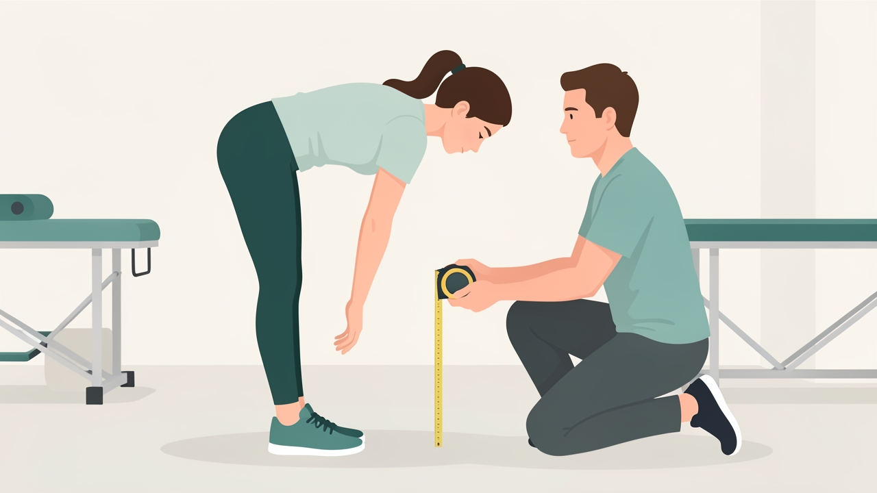
Finger-to-floor distance measures total spinal flexibility: normal is 0 cm (fingers touch the floor), and more than 10 cm indicates significant limitation.
- Flexion: 40–60° (measured from neutral to full forward bend)
- Extension: 20–30°
- Lateral flexion (each side): 15–20°
- Rotation: 5–10° (limited by facet joint orientation)
Schober’s test (modified) measures lumbar flexion: mark 10 cm above and 5 cm below the posterior superior iliac spines (PSIS) in standing; on forward flexion, the distance should increase by at least 5 cm. A value <5 cm suggests hypomobility.
Finger-to-floor distance (FTF) is a simple measure of total spinal flexibility; normal is 0 cm (fingers touch floor). Values >10 cm indicate significant limitation.
Clinical Assessment of Lumbar Anatomy
A systematic assessment of lumbar anatomy should include:
- Observation: Posture, symmetry, muscle atrophy, skin changes.
- Palpation: Spinous processes, paraspinal muscles, PSIS, sacroiliac joints, greater trochanters.
- Active ROM: Flexion, extension, lateral flexion, rotation – record pain location and quality.
- Passive intervertebral motion (PIVM): Assess segmental mobility at each level (L1–S1) using posterior-anterior pressure. Normal is a springy end-feel; hypomobility (stiff) or hypermobility (excessive) can be graded.
- Neurological exam: Sensation (dermatomes), strength (myotomes), reflexes (L4: patellar; S1: Achilles), and special tests (straight leg raise, crossed SLR, femoral nerve tension test).
- Special tests for specific structures:
- Discogenic pain: Centralization with repeated movements (McKenzie)
- Facet pain: Extension-rotation test
- Sacroiliac joint: Cluster of tests (FABER, Gaenslen, compression, distraction)
Grading for SLR: Normal is 70–90° without leg pain. Reproduction of radicular pain below the knee at <60° suggests nerve root irritation (positive test). Crossed SLR (pain in the symptomatic leg when lifting the contralateral leg) has high specificity (>90%) for disc herniation.
Evidence-Based Treatment Approaches and Clinical Reasoning
Understanding anatomy guides treatment selection. Key evidence-based approaches include:
- McKenzie Method (Mechanical Diagnosis and Therapy): Based on directional preference. For discogenic pain, repeated extension often centralizes symptoms (moving pain from leg to spine). This is supported by the anatomical fact that extension reduces posterior disc pressure and may retract nuclear material.
- Stabilization exercises: Target multifidus and transversus abdominis. Research shows that isolated multifidus activation reduces recurrence of low back pain (Hides et al., 2001). Exercises such as the “dead bug” and “bird dog” enhance segmental control.
- Manual therapy: Mobilization of hypomobile segments (e.g., grade III–IV posterior-anterior glides) can improve ROM and reduce pain, likely through neurophysiological mechanisms (gate control, descending inhibition).
- Education: Explaining the anatomy of disc herniation and resorption (often over 6–12 months) reduces fear and improves outcomes.
Clinical reasoning example: A patient with acute central low back pain, worse with sitting and forward bending, better with walking and lying prone – this suggests a discogenic source with a directional preference for extension. The clinician should avoid flexion-based exercises and instead prescribe repeated extension in lying (press-ups).
Clinical Pearls
- The PLL is your friend in disc herniations: Central herniations are often contained by the PLL, whereas posterolateral herniations are not. This explains why central herniations may respond better to conservative care.
- Cauda equina syndrome is a red flag: Always screen for saddle anesthesia and bladder function in any patient with acute back pain and bilateral leg symptoms.
- Multifidus atrophy is reversible: With targeted stabilization training, CSA can increase within 4–6 weeks.
- Don’t forget the iliolumbar ligament: In patients with L5–S1 pain and a history of repetitive twisting, consider iliolumbar ligament sprain.
- Facet joint pain rarely refers below the knee: If leg pain is present, suspect disc or nerve root involvement.
References
Frobin, W., Brinckmann, P., & Biggemann, M. (1997). Precision measurement of disc height, vertebral height and sagittal plane displacement from lateral radiographic views of the lumbar spine. Clinical Biomechanics, 12(3), S1–S61.
Hides, J. A., Richardson, C. A., & Jull, G. A. (1996). Multifidus muscle recovery is not automatic after resolution of acute, first-episode low back pain. Spine, 21(23), 2763–2769.
Hides, J. A., Jull, G. A., & Richardson, C. A. (2001). Long-term effects of specific stabilizing exercises for first-episode low back pain. Spine, 26(11), E243–E248.
Modic, M. T., Steinberg, P. M., Ross, J. S., Masaryk, T. J., & Carter, J. R. (1988). Degenerative disk disease: assessment of changes in vertebral body marrow with MR imaging. Radiology, 166(1), 193–199.
Pfirrmann, C. W., Metzdorf, A., Zanetti, M., Hodler, J., & Boos, N. (2001). Magnetic resonance classification of lumbar intervertebral disc degeneration. Spine, 26(17), 1873–1878.
Verbiest, H. (1954). A radicular syndrome from developmental narrowing of the lumbar vertebral canal. Journal of Bone and Joint Surgery, 36-B(2), 230–237.
Weishaupt, D., Zanetti, M., Boos, N., & Hodler, J. (1999). MR imaging and CT in osteoarthritis of the lumbar facet joints. Skeletal Radiology, 28(4), 215–219.