Introduction
Sensory evaluation is a cornerstone of the neurological examination, providing critical insight into the integrity of the peripheral and central nervous systems. The sensory system encompasses multiple modalities—including touch, pain, temperature, vibration, and proprioception—each mediated by distinct receptor types, peripheral nerve fibers, and central pathways. A thorough understanding of sensory anatomy and physiology is essential for accurate localization of lesions, differentiation of peripheral from central disorders, and formulation of differential diagnoses. This chapter introduces the foundational principles of sensory systems, emphasizing clinically relevant anatomy, examination protocols, normal parameters, and common pathological findings. Mastery of these concepts enables the clinician to perform a focused, efficient, and interpretable sensory assessment.
Anatomy and Physiology of Sensory Systems
Receptor Types and Fiber Classes
Sensory receptors are specialized endings of primary afferent neurons. Mechanoreceptors (Meissner’s corpuscles, Pacinian corpuscles, Merkel cells, Ruffini endings) subserve light touch, pressure, and vibration. Nociceptors respond to noxious stimuli (mechanical, thermal, chemical) and are free nerve endings. Thermoreceptors detect cold (Aδ fibers) and warmth (C fibers). Proprioceptors (muscle spindles, Golgi tendon organs, joint receptors) provide position sense.
Peripheral nerve fibers are classified by diameter and conduction velocity:
- Aα fibers (large, myelinated): proprioception, motor efferents (12–20 μm, 70–120 m/s)
- Aβ fibers (large, myelinated): light touch, vibration (6–12 μm, 30–70 m/s)
- Aδ fibers (small, myelinated): sharp pain, cold (1–5 μm, 5–30 m/s)
- C fibers (unmyelinated): dull pain, warmth, itch (0.2–1.5 μm, 0.5–2 m/s)
Central Pathways
Dorsal column–medial lemniscal pathway (discriminative touch, vibration, proprioception): First-order neurons in dorsal root ganglia (DRG) ascend ipsilaterally in the fasciculus gracilis (lower body) and fasciculus cuneatus (upper body). They synapse in the nucleus gracilis and cuneatus of the medulla. Second-order neurons decussate as internal arcuate fibers and form the medial lemniscus, ascending to the ventral posterolateral (VPL) nucleus of the thalamus. Third-order neurons project to the primary somatosensory cortex (postcentral gyrus).
Spinothalamic tract (pain, temperature, crude touch): First-order neurons in DRG synapse in the substantia gelatinosa (lamina II) of the dorsal horn. Second-order neurons decussate within one to two spinal segments via the anterior white commissure and ascend in the lateral spinothalamic tract (pain, temperature) and anterior spinothalamic tract (crude touch). They terminate in the VPL nucleus; third-order neurons project to the cortex.
Dorsal spinocerebellar tract carries unconscious proprioception from muscle spindles and Golgi tendon organs to the cerebellum for coordination.
Clinical Examination Techniques
General Principles
The sensory examination requires patient cooperation, alertness, and consistent instructions. Test each modality in a systematic order, comparing symmetric areas on both sides, and document the dermatomal or peripheral nerve distribution of any abnormality. Begin with the least noxious stimuli. Use a quiet, well-lit room. The patient should be comfortable and able to see the stimulus (except when testing eyes-closed modalities). Always test with eyes closed for discriminative modalities (vibration, proprioception, stereognosis) to avoid visual cues.
Step-by-Step Protocol
Light Touch
Use a cotton wisp or a fine brush. Apply gentle, brief strokes on the skin. Ask the patient to say "yes" each time they feel the touch. Test all dermatomes from C2 to S5, covering face, trunk, and limbs. Record areas of hypoesthesia or hyperesthesia.
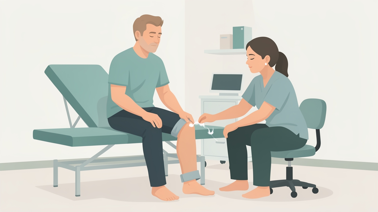
Light touch: brief, gentle strokes with a cotton wisp. The patient says "yes" each time they feel it, and symmetric areas are compared on both sides.
Pain (Pinprick)
Use a sterile safety pin or a neurotip. Alternate sharp and dull ends. Ask the patient to differentiate "sharp" from "dull." Do not use excessive pressure. Test dermatomes similarly. Note hyperalgesia (increased pain response), hypoalgesia (reduced pain), or allodynia (pain from normally non-painful stimulus).
Temperature
Use a metal tuning fork cooled in cold water (cold) and a tube of warm water (40–45°C). Apply briefly. Ask patient to identify "cold" or "warm." Test over dermatomes. Because pain and temperature travel together in the spinothalamic tract, isolated temperature loss suggests a central lesion.
Vibration
Use a 128 Hz tuning fork. Place the base on a bony prominence (e.g., distal interphalangeal joint of index finger, great toe, malleoli). Ask patient to indicate when vibration stops. Compare with examiner’s own sensation (assuming normal). Normal vibration sense declines with age; a rough guide: perception at great toe should last >10 seconds in young adults, >5 seconds in elderly.
Proprioception (Position Sense)
Hold the patient’s distal phalanx of the index finger or great toe by the sides. Move the digit up or down about 5–10 mm. Ask patient to identify direction while eyes closed. Test at distal interphalangeal joints, then proximal joints if distal is impaired. Normal: correct identification of small movements (1–2 mm at finger, 5 mm at toe).
Additional Tests
- Stereognosis: Place a familiar object (e.g., key, coin) in the patient’s hand; ask to identify with eyes closed.
- Graphesthesia: Trace a number (e.g., 3, 5) on the palm; ask patient to identify.
- Two-point discrimination: Use a caliper or paper clip; normal values: finger pulp 2–4 mm, palm 8–12 mm, back 40–60 mm.
Normal Values and Parameters
- Light touch: Perception of a cotton wisp is normal across all dermatomes.
- Pinprick: Clear discrimination of sharp vs. dull; no allodynia.
- Temperature: Able to differentiate cold (20–25°C) from warm (40–45°C) within 2–3 seconds.
- Vibration: Perception of 128 Hz tuning fork at great toe for at least 10 seconds in adults <60 years. Age-adjusted: subtract 1 second per decade over 60.
- Proprioception: Correct identification of movement as small as 1–2 mm at fingers, 5 mm at toes.
- Two-point discrimination: Varies by body region; fingertips most sensitive (2–4 mm).
Common Pathological Findings and Their Clinical Significance
Peripheral Neuropathy
- Length-dependent pattern (stocking-glove): impaired vibration and proprioception first (large fiber), then pain/temperature (small fiber). Common in diabetes, alcoholism, chemotherapy.
- Mononeuropathy: sensory loss in distribution of a single nerve (e.g., median, ulnar, radial).
Spinal Cord Lesions
- Dorsal column lesion: Ipsilateral loss of vibration and proprioception below the lesion; Romberg sign positive. Seen in tabes dorsalis, vitamin B12 deficiency, multiple sclerosis.
- Spinothalamic tract lesion: Contralateral loss of pain and temperature 1–2 segments below the lesion (Brown-Séquard syndrome). Ipsilateral motor loss and dorsal column loss.
- Central cord lesion: Bilateral loss of pain and temperature in a cape distribution (syringomyelia, intramedullary tumor). Dorsal columns spared.
Brainstem Lesions
- Lateral medullary syndrome (Wallenberg): ipsilateral facial pain/temperature loss (trigeminal nucleus), contralateral body pain/temperature loss (spinothalamic tract), ipsilateral Horner’s, ataxia.
- Medial medullary syndrome: contralateral loss of vibration and proprioception (medial lemniscus), contralateral hemiparesis (corticospinal), ipsilateral tongue weakness.
Thalamic and Cortical Lesions
- Thalamic stroke: contralateral hemisensory loss (all modalities), often with severe pain (thalamic pain syndrome, Dejerine-Roussy).
- Cortical lesion (parietal lobe): impaired discriminative modalities (stereognosis, graphesthesia, two-point discrimination) with preserved primary sensation. Sensory neglect, extinction on double simultaneous stimulation.

Thalamic stroke: contralateral hemisensory loss in all modalities, often with severe pain (thalamic pain syndrome, Dejerine-Roussy).
Differential Diagnosis Considerations
When a patient presents with sensory symptoms, consider the following patterns:
- Polyneuropathy: Distal symmetric, length-dependent. Check for diabetes, B12, thyroid, toxins.
- Radiculopathy: Dermatomal distribution, often with pain. Check for herniated disc, herpes zoster.
- Plexopathy: Multiple peripheral nerves in one limb. Consider trauma, tumor, inflammatory (Parsonage-Turner).
- Mononeuropathy: Single nerve distribution. Look for entrapment (carpal tunnel, ulnar at elbow).
- Myelopathy: Sensory level, spasticity, bowel/bladder involvement. MRI spine indicated.
- Brainstem or thalamic: Crossed findings (face vs. body) or hemisensory loss.
- Functional sensory loss: Non-anatomic distribution, inconsistency, give-way weakness, Hoover sign. Use careful examination techniques.
Clinical Pearls and Evidence-Based Recommendations
- Always test pain and temperature separately – isolated loss of one suggests central lesion; combined loss suggests peripheral or spinal cord.
- Use a 128 Hz tuning fork for vibration – 256 Hz is less reliable; 128 Hz has been validated in clinical studies (O’Brien, 2005).
- Proprioception should be tested at the most distal joint – if intact, proximal joints are rarely abnormal.
- Romberg test – stand with feet together, eyes open then closed. Positive if sway increases with eyes closed (dorsal column dysfunction) but not cerebellar (sway even with eyes open).
- In elderly patients, mild distal vibration loss is common; compare with age-matched norms and document.
- For suspected small fiber neuropathy, consider skin biopsy for intraepidermal nerve fiber density (IENFD) – gold standard (Lauria et al., 2010).
- Sensory nerve conduction studies can confirm large fiber involvement but are normal in pure small fiber neuropathy.
- Bedside testing of temperature with a cold tuning fork is rapid and reliable; warm water is more sensitive for detecting subtle loss.
References
- Gilman S. Joint position sense and vibration sense: anatomical organisation and assessment. J Neurol Neurosurg Psychiatry. 2002;73(5):473-477. doi:10.1136/jnnp.73.5.473
- O’Brien MD. Aids to the Examination of the Peripheral Nervous System. 5th ed. Saunders; 2010.
- Lauria G, Hsieh ST, Johansson O, et al. European Federation of Neurological Societies/Peripheral Nerve Society Guideline on the use of skin biopsy in the diagnosis of small fiber neuropathy. Eur J Neurol. 2010;17(7):903-912. doi:10.1111/j.1468-1331.2010.03023.x
- Campbell WW. DeJong’s The Neurologic Examination. 8th ed. Wolters Kluwer; 2020.
- Brazis PW, Masdeu JC, Biller J. Localization in Clinical Neurology. 7th ed. Wolters Kluwer; 2016.
- Shy ME, Frohman EM, So YT, et al. Quantitative sensory testing: report of the Therapeutics and Technology Assessment Subcommittee of the American Academy of Neurology. Neurology. 2003;60(6):898-904. doi:10.1212/01.wnl.0000058546.16913.11
- Dyck PJ, O’Brien PC, Kosanke JL, Gillen DA, Karnes JL. A 4, 2, and 1 stepping algorithm for quick and accurate estimation of cutaneous sensation threshold. Neurology. 1993;43(8):1508-1512. doi:10.1212/wnl.43.8.1508