Introduction to Lateral Elbow Pain: A Clinical Overview of Lateral Epicondylalgia
Lateral elbow pain, commonly referred to as tennis elbow or lateral epicondylalgia, represents one of the most prevalent musculoskeletal disorders encountered in clinical practice. This condition, historically misnamed lateral epicondylitis due to the erroneous assumption of an inflammatory process, is now understood to be a degenerative tendinopathy primarily affecting the extensor carpi radialis brevis (ECRB) tendon. The term "lateral epicondylalgia" is preferred as it accurately describes pain without implying a specific pathology. This chapter provides a comprehensive introduction to the epidemiology, demographics, natural history, risk factors, and clinical presentation of lateral epicondylalgia, emphasizing evidence-based clinical reasoning and decision-making for physiotherapists.
Epidemiology and Demographics
Lateral epicondylalgia affects 1–3% of the general population annually, with a lifetime prevalence of up to 50% among tennis players and 15–20% among individuals engaged in repetitive manual occupations. The condition shows a peak incidence between the ages of 40 and 60 years, with no significant sex predilection, although some studies report a slight female predominance. The dominant arm is affected in 70–80% of cases, and bilateral involvement occurs in 10–15% of patients. Among tennis players, the incidence correlates with playing frequency, racket weight, and technique, with recreational players affected more often than elite athletes due to improper mechanics and conditioning.
Occupational risk is substantial: workers in manufacturing, construction, and food processing have prevalence rates of 10–20%, while office workers with repetitive computer mouse use show rates of 5–10%. A landmark epidemiological study by Shiri et al. (2006) reported an annual incidence of 4.2 per 1000 person-years in a Finnish working population. The economic burden is significant, with median time off work ranging from 2 to 6 weeks, and up to 10% of affected workers requiring job modifications or extended leave.
Natural History and Prognosis
The natural history of lateral epicondylalgia is generally favorable, with 80–90% of patients experiencing resolution of symptoms within 12 months with appropriate conservative management. However, recurrence rates are high, ranging from 25% to 50% within two years, particularly if occupational or sports-related risk factors are not addressed. A systematic review by Bisset et al. (2006) found that at one year, 80% of patients reported improvement, but only 50% were completely pain-free. Chronicity is associated with bilateral involvement, high baseline pain intensity, and prolonged duration of symptoms before treatment.
Prognostic factors for poor outcome include: age over 50 years, manual labor occupation, high baseline disability scores on the Patient-Rated Tennis Elbow Evaluation (PRTEE), and presence of concomitant neck pain or cervical radiculopathy. The condition does not typically lead to permanent disability, but persistent pain can significantly impact quality of life, grip strength, and functional activities such as shaking hands, lifting objects, or turning a doorknob.
Risk Factors
Risk factors for lateral epicondylalgia are multifactorial and can be categorized into intrinsic, extrinsic, and occupational/sports-related factors.
Intrinsic factors include age (peak 40–60 years), smoking (odds ratio 2.5), obesity (BMI >30, odds ratio 1.8), diabetes mellitus (odds ratio 2.3), and genetic predisposition (e.g., COL5A1 gene variants). Histological studies show that the ECRB tendon undergoes degenerative changes with aging, including collagen disorganization, mucoid degeneration, and neovascularization, making it more susceptible to overload.
Extrinsic factors involve repetitive mechanical loading. The ECRB tendon is particularly vulnerable due to its anatomical position: it lies directly under the extensor carpi radialis longus (ECRL) and is compressed against the lateral epicondyle during wrist extension and radial deviation. Activities requiring repetitive gripping, wrist extension, or forearm supination—such as using a screwdriver, hammering, typing, or playing tennis—impose tensile and compressive loads exceeding the tendon's capacity. In tennis, risk factors include improper backhand technique, heavy racket head, high string tension, and playing on cold days.
Occupational factors are well-documented: jobs requiring >1 hour/day of forceful gripping or >30 repetitions/minute of wrist extension increase risk by 3–5 times. A dose-response relationship exists: workers performing high-force, high-repetition tasks have an incidence of 15–20%, compared to 2–5% for low-risk jobs.
Clinical Presentation Overview
Patients typically present with insidious onset of pain over the lateral epicondyle, which may radiate into the proximal forearm along the extensor muscle mass. Pain is exacerbated by activities that load the wrist extensors, such as gripping, lifting with the palm down, shaking hands, or extending the wrist against resistance. Night pain is uncommon but may occur if the patient sleeps with the elbow flexed and wrist pronated.
On examination, tenderness is localized to the lateral epicondyle, approximately 1–2 cm distal and anterior to the bony prominence. Key provocative tests include:
- Cozen's test: The patient makes a fist, extends the wrist against resistance, and pain is reproduced over the lateral epicondyle.
- Mills test: The examiner passively flexes the wrist and extends the elbow; pain indicates a positive test.
- Maudsley's test: Resisted extension of the middle finger with the elbow extended provokes pain at the ECRB origin.
These tests have moderate sensitivity (60–80%) and specificity (70–90%) when used in combination. Grip strength is typically reduced by 20–30% compared to the unaffected side, and pain-free grip strength (PFGS) measured with a dynamometer is a reliable outcome measure. The PRTEE is the most commonly used patient-reported outcome measure, assessing pain and function with 15 items scored 0–100 (higher scores = worse disability).
Differential diagnoses must be considered: radial tunnel syndrome (pain more distal over the supinator, positive resisted supination test), posterior interosseous nerve entrapment (weakness of finger extension), intra-articular elbow pathology (loss of motion, catching), and cervical radiculopathy (neck pain, neurological signs). A thorough clinical reasoning approach includes screening the cervical spine, assessing for concomitant tendinopathy (e.g., de Quervain's tenosynovitis), and considering systemic conditions such as rheumatoid arthritis or fibromyalgia.
Pathophysiology and Clinical Reasoning
Current evidence supports a degenerative tendinopathy model. Histopathology reveals angiofibroblastic hyperplasia, collagen fiber disorganization, increased ground substance, and neovascularization—features of failed healing response rather than acute inflammation. Inflammatory cells are notably absent, which explains why anti-inflammatory treatments (e.g., corticosteroid injections) provide only short-term relief and may increase recurrence risk. The term "tendinosis" is more accurate than "tendinitis."
Clinical reasoning for physiotherapy must therefore focus on load management, progressive strengthening, and addressing biomechanical deficits. Imaging is not routinely required but may be used for recalcitrant cases: ultrasound or MRI can show tendon thickening, hypoechoic areas, and tears. However, up to 50% of asymptomatic individuals have abnormal imaging findings, so clinical correlation is essential.
Practical Assessment and Decision-Making
A comprehensive assessment includes:
- History: Onset, duration, aggravating factors, occupation, sports participation, previous treatments.
- Outcome measures: PRTEE, PFGS (normal grip strength 40–50 kg for men, 20–30 kg for women; PFGS <30% of contralateral side suggests significant disability), Numerical Pain Rating Scale (0–10).
- Physical examination: Palpation, active and passive range of motion (elbow extension may be limited by pain), resisted wrist extension and supination, grip strength dynamometry, provocative tests (Cozen, Mills, Maudsley), and cervical spine screening.
- Differential diagnosis: Perform resisted supination test (radial tunnel), resisted middle finger extension (ECRB), and check for neurological signs.
Evidence-based recommendations for assessment include using a cluster of at least three positive tests to increase diagnostic accuracy (sensitivity 85%, specificity 90%). The PRTEE is recommended as the primary patient-reported outcome due to its excellent reliability (ICC 0.89–0.94) and responsiveness.
Evidence-Based Treatment Principles (Overview)
While detailed treatment strategies are covered in subsequent chapters, introductory clinical reasoning emphasizes:
- Load management: Reduce aggravating activities initially, then gradually progressive loading with eccentric exercises (e.g., Tyler twist using a TheraBand FlexBar, 3 sets of 15 repetitions twice daily).
- Manual therapy: Soft tissue mobilization to the forearm extensors, Mulligan mobilization with movement (MWM) for pain relief.
- Modalities: Low-level laser therapy and shockwave therapy have moderate evidence; corticosteroid injections are effective short-term (2–6 weeks) but associated with higher recurrence at 12 months.
- Patient education: Explain the degenerative nature, set realistic expectations (80% improve in 12 months), and address ergonomic modifications (e.g., using a neutral grip, adjusting workstation height).
Clinical Pearls and Practical Tips
- Differentiate from radial tunnel syndrome: Radial tunnel pain is typically 3–5 cm distal to the lateral epicondyle, and the resisted supination test is positive, whereas lateral epicondylalgia is more proximal and positive with wrist extension.
- Use pain-free grip strength as a quick objective measure: A difference of >20% between sides indicates significant impairment; monitor weekly to track progress.
- Don't forget the neck: Up to 15% of patients with lateral elbow pain have a cervical spine component. Always screen for C5–C7 radiculopathy with Spurling's test and upper limb neurodynamic tests.
- Address psychosocial factors: Fear of movement, catastrophizing, and work-related stress can delay recovery. Use the STarT Back tool or similar to identify yellow flags.
- For tennis players: Assess racket grip size (too small increases forearm muscle activity), string tension (lower tension reduces impact), and backhand technique (one-handed backhand increases risk).
Real-World Application Scenarios
Scenario 1: A 45-year-old office worker presents with 3 months of right lateral elbow pain, aggravated by computer mouse use and lifting coffee cups. PRTEE score 48/100, PFGS 18 kg (left 32 kg), positive Cozen and Maudsley tests. Cervical spine screening negative. Management: Ergonomic adjustment (vertical mouse, forearm support), eccentric loading with FlexBar, and manual therapy. Expected improvement: 50% reduction in PRTEE at 6 weeks.
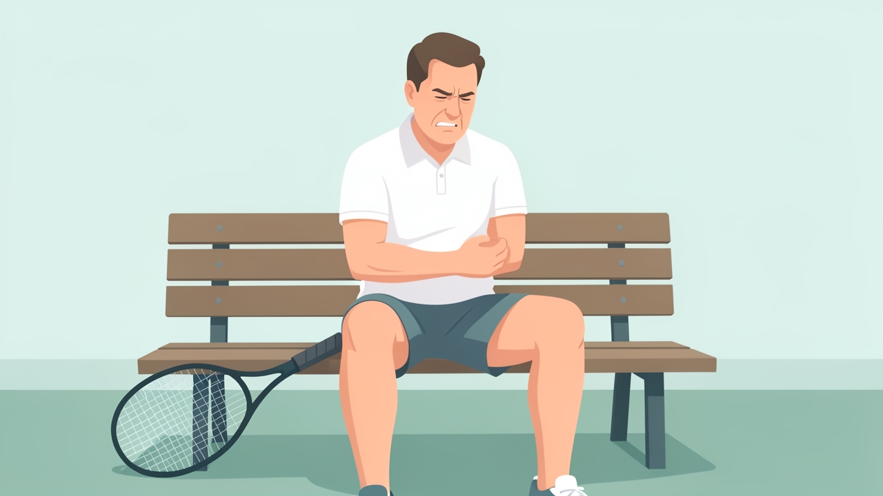
Scenario 2: a 52-year-old recreational tennis player with pain on the backhand and when shaking hands after a weekend tournament.
Scenario 2: A 52-year-old recreational tennis player with acute onset after a weekend tournament. Pain with backhand and shaking hands. PFGS 22 kg (left 40 kg). History of similar episode 2 years ago. Advise relative rest, ice, and gradual return to sport with technique correction (use two-handed backhand). Prescribe eccentric exercises and consider shockwave therapy if no improvement in 4 weeks.
References
- Bisset L, Beller E, Jull G, et al. Mobilisation with movement and exercise, corticosteroid injection, or wait and see for tennis elbow: randomised trial. BMJ. 2006;333(7575):939. doi:10.1136/bmj.38961.584653.AE
- Shiri R, Viikari-Juntura E, Varonen H, Heliövaara M. Prevalence and determinants of lateral and medial epicondylitis: a population study. Am J Epidemiol. 2006;164(11):1065-1074. doi:10.1093/aje/kwj325
- Coombes BK, Bisset L, Vicenzino B. A new integrative model of lateral epicondylalgia. Br J Sports Med. 2009;43(4):252-258. doi:10.1136/bjsm.2008.052738
- Kraushaar BS, Nirschl RP. Tendinosis of the elbow (tennis elbow). Clinical features and findings of histological, immunohistochemical, and electron microscopy studies. J Bone Joint Surg Am. 1999;81(2):259-278. doi:10.2106/00004623-199902000-00014
- Smidt N, van der Windt DA, Assendelft WJ, et al. Corticosteroid injections, physiotherapy, or a wait-and-see policy for lateral epicondylitis: a randomised controlled trial. Lancet. 2002;359(9307):657-662. doi:10.1016/S0140-6736(02)07811-X
- Vicenzino B, Cleland JA, Bisset L. Joint manipulation in the management of lateral epicondylalgia: a clinical commentary. J Man Manip Ther. 2007;15(1):50-56. doi:10.1179/106698107791090150
- Tyler TF, Thomas GC, Nicholas SJ, McHugh MP. Addition of isolated wrist extensor eccentric exercise to standard treatment for chronic lateral epicondylosis: a prospective randomized trial. J Shoulder Elbow Surg. 2010;19(6):917-922. doi:10.1016/j.jse.2010.04.041