1. Hamstring Anatomy and Architecture
The hamstring group comprises three muscles: biceps femoris (long and short heads), semitendinosus, and semimembranosus. All are biarticular, crossing the hip and knee, so they extend the hip and flex the knee. The long head of biceps femoris and semitendinosus share a proximal common origin on the ischial tuberosity; semimembranosus arises from a separate tendon on the same tuberosity. The short head of biceps femoris arises from the lateral lip of the linea aspera and is the only hamstring component without hip action. Its innervation, from the common peroneal division of the sciatic nerve, differs from the tibial-innervated medial hamstrings.

Biceps femoris long head is most vulnerable in terminal swing, lengthened under high eccentric load as the hip flexes and the knee extends.
Architecture matters clinically. Biceps femoris long head has long fascicles (approximately 10 to 12 cm) and is particularly vulnerable during terminal swing, when the muscle is lengthened under high eccentric load while the hip is flexed and the knee is extending. Injury is most common in the long head of biceps femoris, and the proximal musculotendinous junction is a frequent site, particularly in sprinting and stretching-type injuries.
2. Pathophysiology and Injury Mechanisms
Most hamstring strains are non-contact, occurring during high-speed running. The leading mechanism is an eccentric, lengthening contraction in which the muscle is forced to decelerate knee extension while it is stretched at or near its longest length. Mechanical failure is thought to occur at the myotendinous junction, with secondary inflammatory and remodelling changes. Neuromuscular factors, including delayed activation timing, reduced eccentric strength, and altered fatigue responses, are also implicated.
Risk factors that have been consistently associated with injury include prior hamstring strain (the strongest predictor of re-injury), strength deficits or asymmetry, reduced fascicle length, high sprint exposure, and fatigue. Opar and colleagues (2012) provide a comprehensive review of these factors. Hamstring injuries are best understood as multifactorial, with a strong neuromechanical component.
3. Clinical Presentation
Patients typically describe a sudden, sharp pain in the posterior thigh during sprinting, kicking, or stretching, often with a sensation of a "pop" or "tearing." Pain is usually reproduced with active knee extension against resistance and with stretching, particularly with the hip flexed and the knee extended. Gait may be affected, and patients often report difficulty with stair climbing, sitting from standing, and bending.
Proximal injuries commonly cause buttock or sitting pain, which is characteristically worse during prolonged sitting. Ecchymosis may appear within 24 to 72 hours and can migrate distally along the posterior thigh or calf due to gravity. The Pollock et al. (2014) British Athletics classification uses a structured clinical and imaging framework to grade injury location and severity, and it is useful for communicating prognosis.
Red flags requiring escalation:
- Palpable defect or gap at the musculotendinous or tendinous region
- Immediate, severe loss of function with inability to bear weight
- Neurological signs: foot drop, sensory loss in the tibial or common peroneal distribution, or progressive weakness
- Suspected apophyseal avulsion of the ischial tuberosity in skeletally immature athletes (typically under 18), which warrants radiography
- Unilateral calf swelling, tenderness, or erythema, which raises the possibility of deep vein thrombosis and requires medical assessment before any loading or manual therapy
- Systemic symptoms or unexplained posterior thigh pain not consistent with mechanical injury
4. Assessment
Subjective examination: Mechanism of injury, sensation at onset (pop, tearing), prior hamstring history, training load, and sprint exposure. Use the Patient-Reported Outcome Measures: the Lower Extremity Functional Scale (LEFS, range 0 to 80) and, where appropriate, the Hamstring-specific Pain and Function questionnaires used in research.
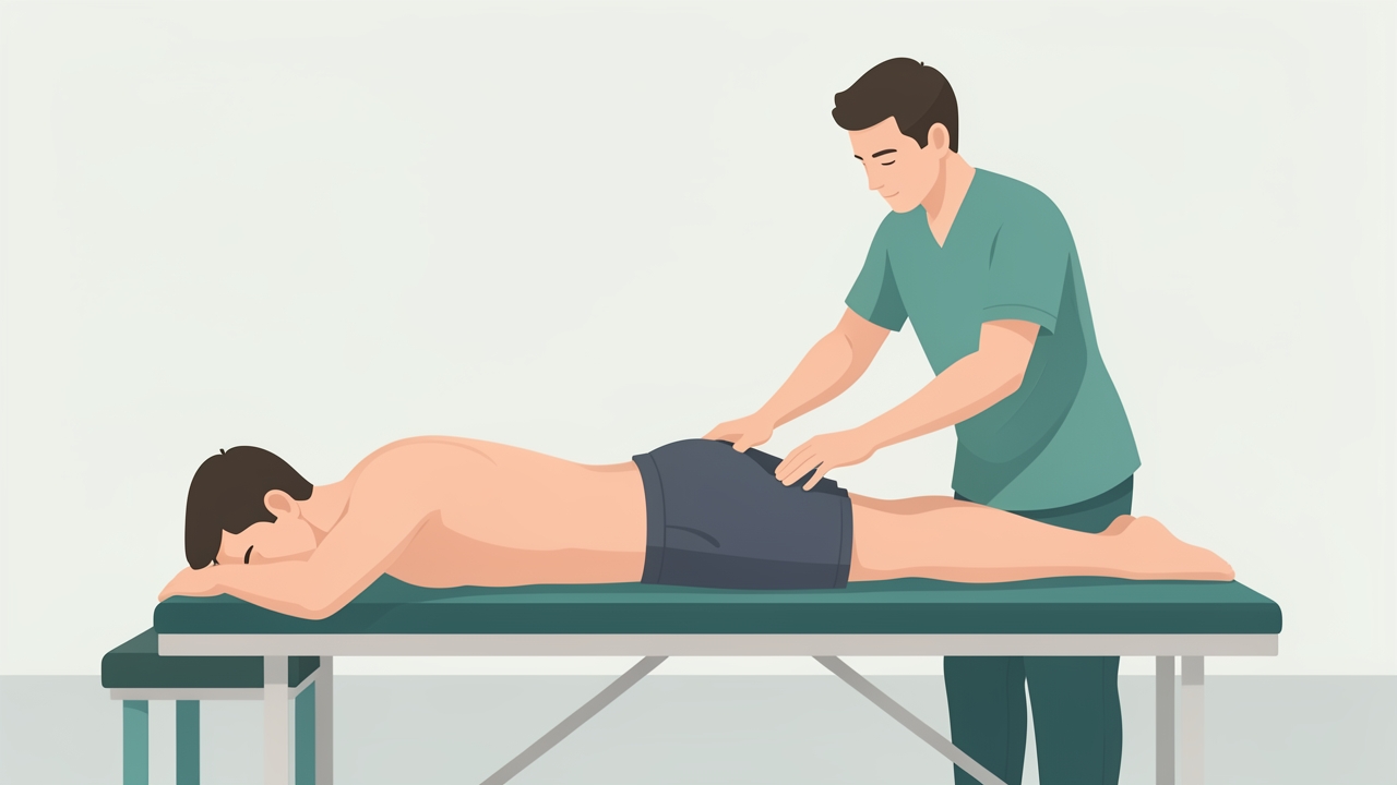
Palpate the ischial tuberosity, proximal musculotendinous junction and mid-belly. Belly palpation with the knee at 90° and the hip neutral helps localise the lesion.
Observation and palpation: Look for gait deviations, ecchymosis, and swelling. Palpate the ischial tuberosity, proximal musculotendinous junction, and mid-belly, noting the location of maximal tenderness. Palpation of the muscle belly with the knee at 90 degrees and the hip neutral helps localise the lesion.
Strength testing:
- Manual muscle testing of knee flexion in prone at 30 degrees of knee flexion (grade 0 to 5)
- Isometric dynamometry at 30 degrees of knee flexion, with the hamstring-to-quadriceps ratio calculated
- Isokinetic dynamometry at 60 degrees per second. Conventional concentric H:Q ratio is typically around 0.6, with values below 0.5 suggesting a strength imbalance
- The Nordic hamstring exercise, measured with a device such as the NordBord, provides eccentric force. Inter-limb asymmetry above roughly 10 to 15 percent is commonly flagged as a risk indicator
Range of motion and flexibility:
- Active knee extension test with hip at 90 degrees. Within 20 degrees of the contralateral side is typical, and a difference greater than 10 to 15 degrees warrants attention
- Straight leg raise to symptom onset, and the Slump test to differentiate neural tension
Functional and performance measures:
- Single-leg hop for distance (limb symmetry index target above 90 percent)
- Askling H-test, a progressive eccentric-loaded test of hamstring flexibility under load
- Sprint testing at maximal velocity, with a 30-metre sprint time compared against baseline
Imaging: Ultrasound or MRI is recommended when the clinical picture is unclear, when a defect is suspected, or when the athlete is considering a return to competitive sport. Imaging should not be used to guide return-to-play decisions alone.
5. Physiotherapy Management: Staged, Graded Protocol
The following staged framework is adapted from Heiderscheit et al. (2010) and Bourne et al. (2018). Timeframes are approximate, and progression should be criterion-based rather than time-based.

Phase 0 (days 0–3): relative rest and pain-free range, no aggressive stretching or sprinting, and crutches for severe gait disturbance.
Phase 0: Protection (days 0 to 3)
- Relative rest, load management, and pain-free range of motion
- Avoid aggressive stretching and sprinting
- Use crutches for severe gait disturbance
- Gentle isometric knee flexion at low intensity (below 30 percent of maximum voluntary contraction) once tolerated
Phase 1: Early loading (days 3 to 14)
- Isometric knee flexion at 30 to 50 percent of maximum, 5 x 45-second holds, once daily or twice daily
- Bridging and prone knee curls with pain-monitoring model: pain should not exceed 3/10 and should return to baseline within 24 hours
- Short-range eccentric training begins once isometrics are pain-free
- Stationary cycling and walking as tolerated
Phase 2: Progressive strengthening (weeks 2 to 6)
- Progress to isotonic knee flexion (3 to 4 sets of 6 to 10 repetitions), 3 times per week with at least 48 hours between sessions
- Introduce the Nordic hamstring exercise, beginning with partial-range or assisted variations (3 sets of 3 to 5 repetitions), progressing toward full range
- Single-leg Romanian deadlift and hip extension exercises under progressive load
- Introduce lengthened-position strengthening, as eccentric training at long muscle lengths has the strongest evidence for hypertrophy and fascicle adaptation
- Begin light jogging when pain-free during and after activity
Phase 3: Return to sport preparation (weeks 6 onwards)
- Progressive sprint exposure: graded acceleration, then maximal velocity sprints with 60 to 80 percent intensity progressing to full speed
- Plyometric training (bounding, hopping) and change-of-direction drills
- Return-to-sport criteria should include: greater than 90 percent limb symmetry on strength and hop tests, pain-free full-speed sprinting, and completion of sport-specific drills without symptoms
Prevention dosage: Nordic hamstring exercise, 2 to 3 sessions per week, 3 sets of 5 to 8 repetitions, integrated into a warm-up or strength programme. Van der Horst et al. (2015) reported a substantial reduction in hamstring injury incidence in amateur football players using this approach.
6. Evidence and Clinical Pearls
Eccentric strengthening, particularly the Nordic hamstring exercise, has the strongest evidence for reducing hamstring injury recurrence and incidence. Bourne et al. (2018) recommend exercises that load the hamstrings at long muscle lengths, such as the Nordic hamstring exercise and Romanian deadlift variants.
Stretching alone has limited evidence for injury prevention, though flexibility is a component of a comprehensive programme. Askling et al. (2007) highlighted the importance of high-speed running in first-time injuries, which suggests that controlled sprint exposure should be part of both rehabilitation and prevention.
Clinical pearls:
- Re-injury risk is highest in the first few weeks after return to sport. Do not discharge based on pain resolution alone.
- Do not clear an athlete based on a single test. Use a battery of strength, flexibility, and performance measures.
- Proximal avulsion and complete tears require surgical opinion, particularly when there is significant retraction or functional deficit.
- Address trunk, pelvic, and lower limb load management, as these influence hamstring demand.
- Screen for lumbar and neural contributions in cases of posterior thigh pain without clear mechanical signs.
7. Summary
Hamstring injuries are multifactorial, with eccentric loading at long muscle lengths, prior injury, and neuromuscular and strength deficits as key contributors. A thorough assessment should include subjective history, palpation, strength and asymmetry measurement, flexibility, and performance testing. Management should follow a criterion-based, staged progression from protection through early isometric loading, progressive eccentric strengthening, and sprint-specific training. Return to sport should be guided by objective symmetry thresholds and pain-free full-speed running, and prevention programmes should include the Nordic hamstring exercise.
References
- Askling, C. M., Tengvar, M., Saartok, T., & Thorstensson, A. (2007). Acute first-time hamstring strains during high-speed running: a longitudinal study including clinical and magnetic resonance imaging findings. American Journal of Sports Medicine, 35(2), 197–206.
- Bourne, M. N., Timmins, R. G., Opar, D. A., et al. (2018). An evidence-based framework for strengthening exercises to prevent hamstring injury. Sports Medicine, 48(2), 251–267.
- Heiderscheit, B. C., Sherry, M. A., Silder, A., Chumanov, E. S., & Thelen, D. G. (2010). Hamstring strain injuries: recommendations for diagnosis, rehabilitation, and injury prevention. Journal of Orthopaedic & Sports Physical Therapy, 40(2), 67–81.
- Mendiguchia, J., Alentorn-Geli, E., & Brughelli, M. (2012). Hamstring strain injuries: are we heading in the right direction? British Journal of Sports Medicine, 46(2), 81–85.
- Opar, D. A., Williams, M. D., & Shield, A. J. (2012). Hamstring strain injuries: factors that lead to injury and re-injury. Sports Medicine, 42(3), 209–226.
- Pollock, N., James, S. L. J., Lee, J. C., & Chakraverty, R. (2014). British athletics muscle injury classification: a new grading system. British Journal of Sports Medicine, 48(18), 1347–1351.
- van der Horst, N., Smits, D. W., Petersen, J., Goedhart, E. A., & Backx, F. J. G. (2015). The preventive effect of the Nordic hamstring exercise on hamstring injuries in amateur soccer players: a randomized controlled trial. American Journal of Sports Medicine, 43(6), 1316–1323.