Introduction
The shoulder complex is one of the most mobile and functionally versatile regions of the human body, enabling a wide range of movements essential for daily activities, sports, and occupational tasks. This chapter provides a comprehensive overview of shoulder anatomy and functional anatomy, integrating clinical details, measurement parameters, assessment procedures, and clinical reasoning to guide physiotherapy practice. Understanding the intricate interplay between bones, joints, muscles, and neural control is critical for accurate diagnosis, effective treatment planning, and prevention of shoulder pathologies.
Osteology
The shoulder girdle consists of three bones: the clavicle, scapula, and proximal humerus. The clavicle acts as a strut connecting the axial skeleton to the upper limb, transmitting forces and protecting underlying neurovascular structures. The scapula is a flat, triangular bone with key landmarks—the acromion, coracoid process, glenoid fossa, spine, and medial/lateral borders. The glenoid fossa is shallow and oriented slightly superiorly and anteriorly, providing minimal bony stability. The proximal humerus includes the head, anatomical neck, surgical neck, greater and lesser tuberosities, and intertubercular groove.
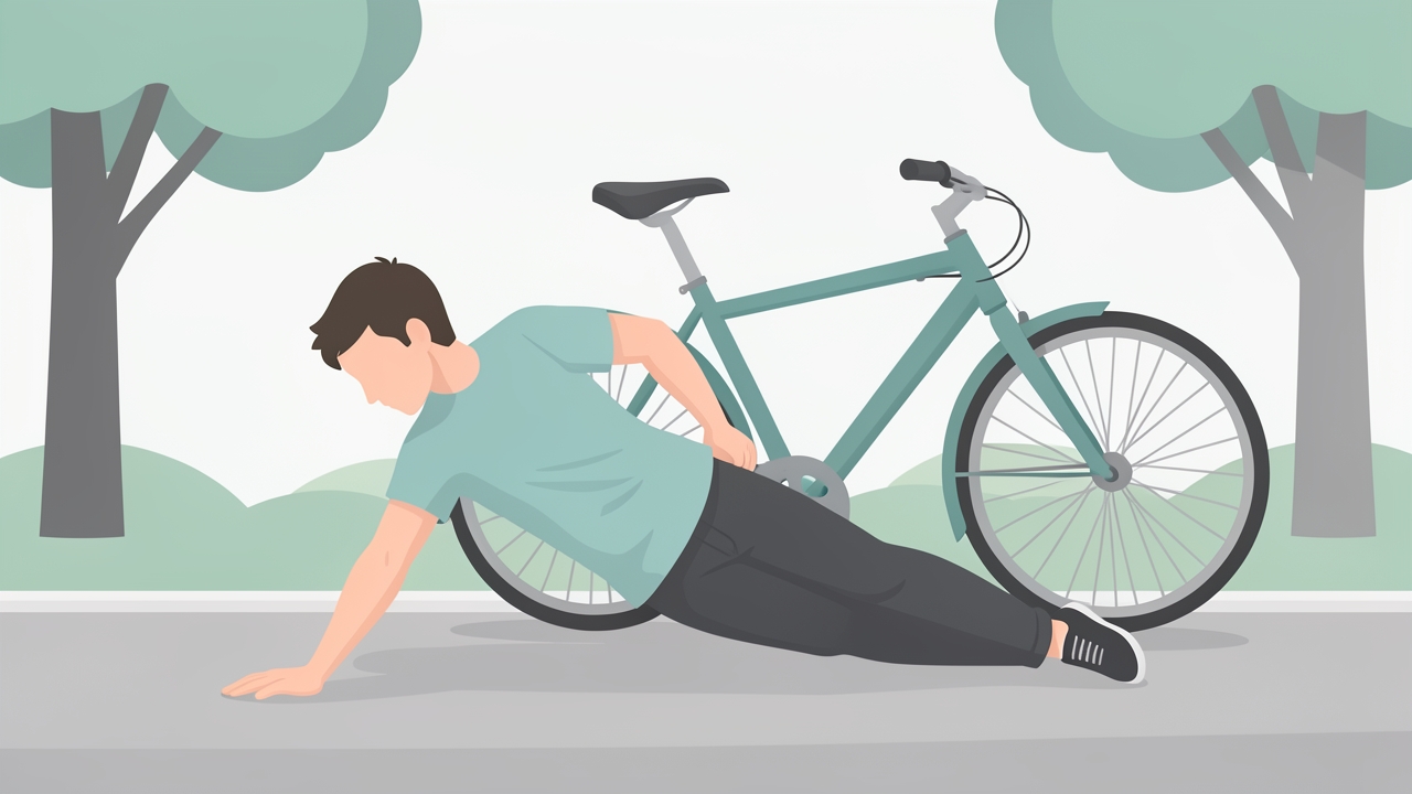
Clavicle fractures are common after a fall onto an outstretched hand (FOOSH); assess for shortening, deformity and neurovascular compromise.
Clinical Relevance
- Clavicle fractures: Common in falls onto an outstretched hand (FOOSH); assess for shortening, deformity, and neurovascular compromise (brachial plexus, subclavian vessels).
- Scapular winging: Medial winging (serratus anterior palsy, long thoracic nerve injury) or lateral winging (trapezius palsy, spinal accessory nerve injury).
- Greater tuberosity fractures: Associated with shoulder dislocations; may cause rotator cuff dysfunction.
- Glenoid dysplasia: Can predispose to recurrent instability.
Arthrology
The shoulder complex comprises four articulations: glenohumeral (GH), acromioclavicular (AC), sternoclavicular (SC), and scapulothoracic (ST).
Glenohumeral Joint
- Type: Ball-and-socket synovial joint.
- Stability: Primarily dependent on dynamic stabilizers (rotator cuff, long head of biceps) and static stabilizers (glenoid labrum, capsular ligaments—superior, middle, inferior glenohumeral ligaments). The inferior glenohumeral ligament is the primary restraint to anterior translation in abduction and external rotation.
- Capsular pattern: Restriction of external rotation > abduction > internal rotation (typical of adhesive capsulitis).
- Norms: GH joint range of motion—flexion 0–180°, extension 0–60°, abduction 0–180°, external rotation 0–90° (arm at side), internal rotation 0–70° (arm at side).
Acromioclavicular Joint
- Type: Plane synovial joint.
- Ligaments: AC ligament (horizontal stability), coracoclavicular ligaments (conoid and trapezoid—vertical stability).
- Clinical tests: AC joint compression test, cross-body adduction test. Pain at AC joint suggests osteoarthritis, separation, or distal clavicle osteolysis.
Sternoclavicular Joint
- Type: Saddle synovial joint (only bony articulation between upper limb and axial skeleton).
- Ligaments: Costoclavicular, interclavicular, and sternoclavicular ligaments.
- Clinical: Anterior dislocation is common (often from direct blow); posterior dislocation is rare but can compress trachea, esophagus, or great vessels.
Scapulothoracic Articulation
- Type: Functional joint (not true synovial); scapula glides over thoracic cage via serratus anterior and subscapularis bursae.
- Scapulohumeral rhythm: 2:1 ratio of GH to ST motion during abduction (e.g., 120° GH, 60° ST for full 180° abduction). Disruption indicates pathology (e.g., rotator cuff tear, frozen shoulder).
Myology
Muscles of the shoulder complex are organized into local stabilizers (rotator cuff) and global movers (deltoid, pectoralis major, latissimus dorsi, etc.).
Rotator Cuff
- Supraspinatus: Abduction (0–30°), external rotation. Innervation: suprascapular nerve (C5–C6). Test: Empty can test (Jobe's)—patient abducts arms to 90° with internal rotation; weakness or pain indicates tear.
- Infraspinatus: External rotation. Innervation: suprascapular nerve. Test: External rotation lag sign; drop arm test.
- Teres minor: External rotation, adduction. Innervation: axillary nerve (C5–C6). Test: Hornblower's sign.
- Subscapularis: Internal rotation. Innervation: upper and lower subscapular nerves (C5–C6). Test: Lift-off test, belly press test.
Scapular Stabilizers
- Trapezius (upper, middle, lower): Elevation, retraction, depression, upward rotation. Innervation: spinal accessory nerve (CN XI). Test: Shoulder shrug, scapular retraction.
- Serratus anterior: Protraction, upward rotation. Innervation: long thoracic nerve (C5–C7). Test: Push-up plus, wall push; winging indicates weakness.
- Rhomboids (major and minor): Retraction, downward rotation. Innervation: dorsal scapular nerve (C4–C5). Test: Scapular retraction with resistance.
- Levator scapulae: Elevation, downward rotation. Innervation: dorsal scapular nerve (C3–C4).
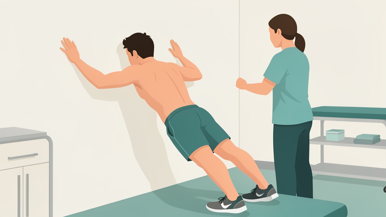
Serratus anterior (long thoracic nerve, C5–C7) is tested with a push-up plus or wall push; winging indicates weakness.
Force Couples
- Scapular force couple: Upper trapezius and lower trapezius with serratus anterior produce upward rotation; levator scapulae and rhomboids produce downward rotation.
- GH force couple: Deltoid (abduction) and rotator cuff (compression) stabilize the humeral head against the glenoid. Imbalance leads to superior migration and impingement.
Functional Anatomy
Scapulohumeral rhythm is essential for efficient overhead motion. During the first 30° of abduction, scapula may "set" (slight elevation or depression) followed by coordinated upward rotation. Kinematically, the humeral head must translate inferiorly and posteriorly to avoid impingement against the acromion. The subacromial space (between humeral head, acromion, and coracoacromial ligament) narrows with abduction, making the supraspinatus tendon vulnerable to compression.
Clinical Reasoning for Impingement
- Neer's test: Passive flexion of the arm (with scapula stabilized) reproduces pain—indicates subacromial impingement.
- Hawkins-Kennedy test: Flexion to 90° with internal rotation—compresses supraspinatus against coracoacromial ligament.
- Empty can test: Assesses supraspinatus integrity; positive if pain or weakness.
Shoulder Instability
- Anterior instability: Most common; apprehension test (abduction + external rotation) elicits fear of dislocation; relocation test (applying posterior force) reduces apprehension.
- Posterior instability: Less common; posterior apprehension test (flexion + adduction + internal rotation).
- Inferior instability: Sulcus sign (inferior translation with traction).
Clinical Assessment Parameters
Range of Motion (Active and Passive)
- Flexion: 0–180° (GH contribution ~120°, ST ~60°).
- Abduction: 0–180° (GH ~120°, ST ~60°).
- External rotation: 0–90° (arm at side); 0–90° (arm abducted 90°).
- Internal rotation: Reach behind back—record vertebral level (e.g., T7). Norm: up to T4–T6.
- Cross-body adduction: 0–30° (AC joint motion).
Manual Muscle Testing (MMT)
- Grade 0–5: Based on ability to move through full ROM against gravity and resistance.
- Supraspinatus: Grade 5 if able to hold abduction at 90° with maximal resistance.
- Infraspinatus: Grade 5 if able to externally rotate against resistance.
- Subscapularis: Grade 5 if able to internally rotate against resistance.
Special Tests
- O'Brien's test (active compression test): For labral tears (SLAP). Patient flexes shoulder to 90°, adducts 10–15°, internally rotates; pain with internal rotation and relief with external rotation suggests labral pathology.
- Speed's test: For biceps tendinopathy; resisted forward flexion with forearm supinated—pain in bicipital groove.
- Yergason's test: Supination with elbow flexed 90°—pain indicates biceps tendon instability.
- Drop arm test: Patient slowly lowers abducted arm; if unable to control, indicates rotator cuff tear.
Neurological Assessment
- Axillary nerve: Deltoid strength; sensation over lateral shoulder (regimental badge area).
- Suprascapular nerve: Supraspinatus/infraspinatus strength.
- Long thoracic nerve: Serratus anterior (winging with push-up).
- Spinal accessory nerve: Trapezius strength.
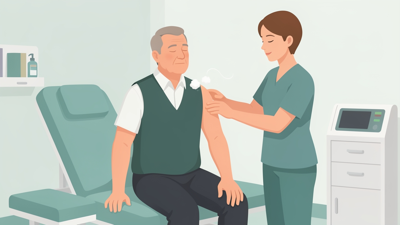
Axillary nerve: check deltoid strength and sensation over the lateral shoulder, the regimental badge area.
References
- Magee DJ. Orthopedic Physical Assessment. 7th ed. Elsevier; 2020.
- Neumann DA. Kinesiology of the Musculoskeletal System: Foundations for Rehabilitation. 3rd ed. Elsevier; 2016.
- Wilk KE, Reinold MM, Andrews JR. The Athlete's Shoulder. 2nd ed. Churchill Livingstone; 2009.
- Ludewig PM, Phadke V, Braman JP, Hassett DR, Cieminski CJ, LaPrade RF. Motion of the shoulder complex during multiplanar humeral elevation. J Bone Joint Surg Am. 2009;91(2):378-389.
- Michener LA, Walsworth MK, Doukas WC, Murphy KP. Reliability and diagnostic accuracy of 5 physical examination tests and combination of tests for subacromial impingement. Arch Phys Med Rehabil. 2009;90(11):1898-1903.
- Hegedus EJ, Goode AP, Cook CE, et al. Which physical examination tests provide clinicians with the most value when examining the shoulder? Update of a systematic review with meta-analysis of individual tests. Br J Sports Med. 2012;46(14):964-978.
- Kibler WB, Sciascia A. The role of the scapula in athletic shoulder function. Am J Sports Med. 2010;38(3):623-633.
- Terry GC, Chopp TM. Functional anatomy of the shoulder. J Athl Train. 2000;35(3):248-255.