Neuroanatomy of Concussion
Concussion is a biomechanically induced brain injury. It results from direct impact or from rotational and translational acceleration-deceleration forces transmitted to the head. Structural imaging is usually normal, so the diagnosis is clinical. Understanding why certain structures are vulnerable helps explain the symptom profile you will assess.
Rotational forces are the strongest predictor of injury severity because they shear white matter tracts. The corpus callosum, brainstem, and deep white matter are especially susceptible. Axons stretched during shear may undergo microstructural change (diffuse axonal injury), even when no macroscopic lesion is visible.
Several functional networks are commonly affected:
- Brainstem and reticular activating system: involvement may contribute to altered arousal, fatigue, and sleep disturbance.
- Vestibular and oculomotor pathways: the vestibulocerebellum, brainstem nuclei, and their connections to the vestibulo-ocular reflex (VOR) and smooth pursuit systems drive dizziness, visual motion sensitivity, and gaze instability.
- Prefrontal and temporal cortex: these regions mediate working memory, processing speed, and attention, which underpin many cognitive complaints in the classroom.
- Cervical afferent system: the upper cervical spine (C1–C3) shares trigeminocervical convergence with the trigeminal nucleus. Neck injury frequently coexists with concussion and can amplify headache and dizziness.
Pathophysiology: The Neurometabolic Cascade
Giza and Hovda's model describes a sequence of events that explains the temporary functional impairment of concussion, even without structural damage. The cascade has several interacting phases.
Phase 1: Ionic flux. Mechanical deformation triggers a massive efflux of potassium (K⁺) and influx of sodium (Na⁺) and calcium (Ca²⁺). Excess extracellular glutamate amplifies this depolarization.
Phase 2: Energy demand. Restoring ionic gradients requires large amounts of adenosine triphosphate (ATP) via Na⁺/K⁺-ATPase activity. Glucose uptake and glycolysis increase sharply, producing a state of hypermetabolism in the injured tissue.
Phase 3: Hemodynamic mismatch. Cerebral blood flow falls in the acute and subacute period, while metabolic demand is elevated. The result is a relative energy crisis. Calcium overload may also impair mitochondrial function, reducing oxidative phosphorylation and shifting metabolism toward anaerobic pathways with lactate accumulation.
Phase 4: Recovery. Ionic homeostasis and cerebral blood flow gradually normalize over days to weeks. Clinical recovery usually lags behind metabolic recovery in many athletes, which is one rationale for graded rather than immediate return to full demand.
For physiotherapists, the practical implication is that the brain is in a vulnerable, energy-limited state. Exertion, cognitive load, and sensory stimulation may exceed available reserve and provoke symptoms, which is why graded, symptom-guided progression is preferred over both complete rest and unrestricted activity.
Clinical Presentation
Symptoms typically fall into four domains:
- Somatic: headache, dizziness, nausea, visual disturbance, fatigue, and sensitivity to light or noise.
- Cognitive: slowed processing, difficulty concentrating, memory lapses, and "fogginess."
- Emotional: irritability, anxiety, low mood, and emotional lability.
- Sleep: insomnia, hypersomnia, or disrupted sleep architecture.
Symptoms may be delayed by hours or even days. Loss of consciousness occurs in fewer than 10% of concussions, and its absence does not exclude injury.
Red flags requiring urgent medical referral:
- Decreasing level of consciousness, or Glasgow Coma Scale (GCS) below 15 at any point
- Repeated vomiting or worsening headache
- Seizure activity
- Pupillary asymmetry or focal neurological deficit
- Progressive confusion, agitation, or slurred speech
- Severe neck pain, midline cervical tenderness, or new neurological signs in the limbs (suspected cervical spine injury)
- Suspected skull fracture or cerebrospinal fluid leakage
Any red flag takes priority over the remainder of the assessment. Athletes should not be left alone and should be transported for emergency assessment.
Assessment: Core Tools and Normative Values
The Sport Concussion Assessment Tool, 5th edition (SCAT5) is the consensus-endorsed screening instrument for athletes aged 13 years and older. Its key components include:
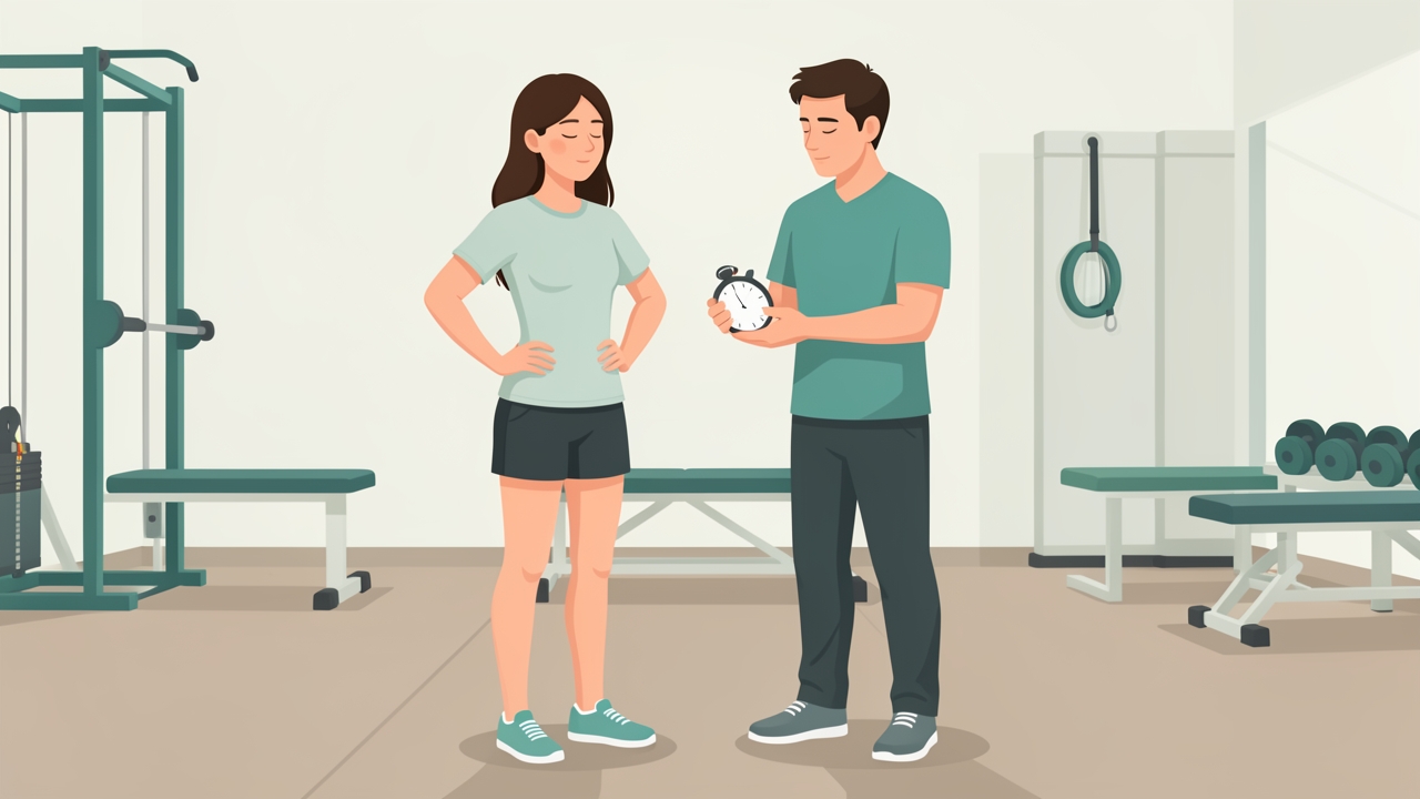
BESS: double-leg, single-leg and tandem stances on firm and foam surfaces, 20 seconds each. Errors are counted, up to 10 per condition.
- Symptom Evaluation: 22 symptoms rated 0–6. Total symptom severity score ranges 0–132. Compare against the athlete's pre-injury baseline wherever possible.
- Glasgow Coma Scale: 15 is normal.
- Cognitive screening: Orientation, immediate memory (five-word lists over three trials), and delayed recall after five minutes. Maximum score on the immediate memory section is 15.
- Balance Error Scoring System (BESS): double-leg, single-leg (non-dominant), and tandem stance on firm and foam surfaces, each for 20 seconds. Errors are counted, with a maximum of 10 per condition. Interpret against age-specific and baseline values, as normative data vary by population.
- Coordination and tandem gait: scored for speed and accuracy.
The Child SCAT5 (ages 5–12) and Sport Concussion Assessment Tool for Children versions adapt these measures to developmental level.
Symptom-based monitoring: The Post-Concussion Symptom Scale (PCSS) and visual analogue scales (0–10) are used to track symptoms across the rehabilitation period. A commonly applied clinical rule is that an increase of more than 2 points above baseline on a symptom during or after exertion indicates the activity is too demanding.
Oculomotor and vestibular screening: The Vestibular/Ocular Motor Screening (VOMS) tool assesses smooth pursuits, saccades, near-point convergence, horizontal and vertical VOR, visual motion sensitivity, and the dizziness rating. Near-point convergence distance greater than 5 cm, or symptom reproduction with VOR testing, indicates a need for targeted intervention. The Dizziness Handicap Inventory (DHI) provides a patient-reported measure, with higher scores indicating greater disability.
Cervical assessment: Assess active range of motion, the Neck Disability Index (NDI), and cervical flexion-rotation test (CFRT). Side-to-side rotational asymmetry, particularly in the upper cervical spine, is commonly flagged at around 10° or with symptom reproduction. Deep neck flexor endurance (craniocervical flexion test) and cervical proprioception (joint position error) should also be recorded.
Exertional testing: The Buffalo Concussion Treadmill Test or a symptom-limited graded aerobic test is used to determine the exertional threshold. Heart rate is monitored, and the test is stopped at symptom onset. Many clinicians use approximately 80% of age-predicted maximum heart rate (220 minus age) as an initial exertional target in the early phase, ensuring the athlete remains below symptom threshold.
Physiotherapy Management: Acute Phase
Management begins with education, reassurance, and a brief period of relative rest. The Berlin consensus (McCrory et al., 2017) supports a short period of rest, typically 24–48 hours, followed by progressive, symptom-guided activity. Prolonged strict rest is no longer recommended, as it is associated with deconditioning, mood disturbance, and delayed recovery.
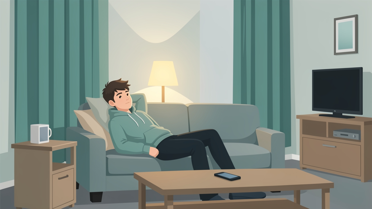
A short period of relative rest, typically 24–48 hours: limit screens, bright light and loud environments without stopping daily function.
Key principles in the acute phase:
- Limit screen time, bright lighting, and loud environments without eliminating daily function.
- Encourage light walking and routine activities of daily living as tolerated.
- Avoid high-risk activities, including contact sport and activities with fall risk, until cleared.
- Monitor symptoms and sleep. Advise against alcohol and non-prescribed sedatives.
- Refer for medical review if any red flag is present or if symptoms are worsening.
Graded Exertion and Return-to-Sport Progression
The Berlin 2016 consensus outlines a six-stage return-to-play protocol. Each stage typically lasts a minimum of 24 hours. Progression proceeds only if the athlete remains symptom-free or within the 2-point threshold during and after activity. If symptoms recur, the athlete returns to the previous stage.
- Symptom-limited activity: daily activities at a level that does not provoke worsening symptoms.
- Light aerobic exercise: walking or stationary cycling for 10–20 minutes at low intensity, below symptom threshold.
- Sport-specific exercise: running or skating drills, with no head impact. Increase intensity and duration gradually.
- Non-contact training drills: more complex, higher-intensity training, with coordination and cognitive load.
- Full-contact practice: after medical clearance, with normal training activities.
- Return to competition.
Vestibular and oculomotor rehabilitation is indicated when screening shows persistent dizziness, visual motion sensitivity, or convergence insufficiency. Typical prescriptions include:
- Gaze stabilization (VOR adaptation): 2–3 sessions per day, 1–2 minutes per exercise, progressing from stationary targets to head movement against a target.
- Smooth pursuit and saccade training: 5–10 minutes daily, progressing to complex visual backgrounds.
- Habituation exercises: head movements in the provoking plane to tolerance, typically 1–2 minutes per exercise, with a brief rest between repetitions.
- Vestibular rehabilitation should be symptom-guided, with a mild, temporary increase in dizziness acceptable but resolving within 30 minutes.
Cervical rehabilitation: Early-phase cervical treatment focuses on pain-free active range, joint position sense, and deep neck flexor activation (craniocervical flexion, 10 repetitions of 10-second holds). Progress to isometric neck strengthening in all directions, initially at low load (for example, 30% of maximum voluntary contraction) and increasing to 3 sets of 10–15 repetitions as tolerated. Manual therapy to the upper cervical spine may be used when cervicogenic contributors are identified, with consideration of contraindications such as suspected vertebrobasilar insufficiency, instability, or unresolved red flags.
Return to Learning
Cognitive exertion can provoke symptoms in the same way as physical activity. Return to learning should be planned alongside, and often before, return to sport.

Return to learning starts with a reduced academic load, such as half days or shortened periods, plus scheduled rest breaks.
A graded approach includes:
- Reduced academic load initially, such as half days or shortened periods.
- Scheduled rest breaks during the day, and reduced screen exposure.
- Accommodations such as extra time for tests, note-taking support, and reduced homework.
- Gradual reintegration toward full timetable, with symptom monitoring at each stage.
Schools should be involved through a documented individualized plan, with clear communication between the clinician, family, and educators.
Evidence Base and Clinical Pearls
Most athletes recover within 7–10 days, though a substantial minority have symptoms persisting beyond 4 weeks. Persistent symptoms beyond this point should prompt review for contributing factors, including vestibular, oculomotor, cervical, mood, and sleep disorders (Ellis et al., 2016). Exercise is an effective and safe intervention for most athletes once the acute phase has passed (Leddy et al., 2018).
Clinical pearls:
- Baseline testing matters. Where pre-season baseline data exist, they provide far more meaningful comparisons than population norms.
- Symptoms guide progression, not time alone. A calendar-based return is less reliable than a symptom-free progression.
- Screen for the neck and vestibular system in every case. Many persistent symptoms have peripheral contributors that are treatable.
- Document everything. Clear records of symptom scores, stage progression, and clearance decisions protect the athlete and the clinician.
- Watch for mood and sleep. Anxiety, depression, and insomnia commonly prolong recovery and should be addressed proactively.
Summary
Concussion is a functional brain injury arising from biomechanical forces, with a neurometabolic cascade producing a temporary energy mismatch. Clinical presentation spans somatic, cognitive, emotional, and sleep domains, and red flags must be excluded first. Assessment combines SCAT5, VOMS, cervical, and exertional testing against individual baseline data. Management involves brief relative rest, early symptom-guided activity, a six-stage return-to-play protocol, targeted vestibular and cervical rehabilitation, and a structured return to learning. Progression should be determined by symptoms and objective function rather than time alone.
References
-
McCrory P, Meeuwisse W, Dvořák J, et al. Consensus statement on concussion in sport: the 5th international conference on concussion in sport held in Berlin, October 2016. Br J Sports Med. 2017;51(11):838–847.
-
Giza CC, Hovda DA. The neurometabolic cascade of concussion. J Athl Train. 2001;36(3):228–235.
-
Harmon KG, Drezner JA, Gammons M, et al. American Medical Society for Sports Medicine position statement: concussion in sport. Br J Sports Med. 2013;47(1):15–26.
-
Echemendia RJ, Meeuwisse W, McCrory P, et al. The Sport Concussion Assessment Tool 5th Edition (SCAT5). Br J Sports Med. 2017;51(11):848–850.
-
Ellis MJ, Leddy J, Willer B. Physiological, vestibulo-ocular and cervicogenic post-concussion disorders: an evidence-based classification system with directions for treatment. Brain Inj. 2016;30(4):405–414.
-
Leddy JJ, Haider MN, Ellis MJ, Willer BS. Exercise is medicine for concussion. Curr Sports Med Rep. 2018;17(8):262–270.
-
Schneider KJ, Leddy JJ, Guskiewicz KM, et al. Rest and treatment/rehabilitation following sport-related concussion: a systematic review. Br J Sports Med. 2017;51(12):930–934.
-
Majerske CW, Mihalik JP, Ren D, et al. Concussion in sports: postconcussive activity levels, symptoms, and neurocognitive performance. J Athl Train. 2008;43(3):265–274.