Scapular Thoracic Care for Overhead Athletes

Scapular Thoracic Care for Overhead Athletes

Advanced · 12 chapters · 144 MCQs · 144 flashcards · Illustrated

Advanced assessment and treatment strategies for scapular dyskinesis in throwing and overhead sports

What you'll learn

  1. Chapter 1: Introduction to Scapulothoracic Anatomy

    The scapulothoracic articulation is a functional joint critical to the efficient and healthy performance of overhead athletes.

  2. Chapter 2: Scapular Kinematics and Biomechanics

    The scapula serves as the critical link between the axial skeleton and the upper extremity, enabling efficient force transfer during overhead athletic activities.

  3. Chapter 3: Scapular Dyskinesis Assessment

    Scapular dyskinesis—an alteration in the normal position or motion of the scapula during coupled scapulohumeral movements—is a common finding in overhead athletes.

  4. Chapter 4: Muscle Function and Imbalances

  5. Chapter 5: Thoracic Spine and Rib Cage Influence

  6. Chapter 6: Overhead Throwing Mechanics

    Overhead throwing is a complex, high-velocity movement that places extreme demands on the scapulothoracic joint.

  7. Chapter 7: Swimming and Racquet Sport Demands

  8. Chapter 8: Clinical Examination Techniques

    A comprehensive clinical examination of the scapulothoracic region is essential for identifying the underlying causes of shoulder dysfunction in overhead athletes.

  9. Chapter 9: Rehabilitation Strategies

    Rehabilitation of the scapulothoracic region in overhead athletes requires a systematic, evidence-based approach that addresses both local scapular mechanics and global kinetic chain function.

  10. Chapter 10: Return to Sport Criteria

  11. Chapter 11: Special Populations and Considerations

    Scapulothoracic rehabilitation for overhead athletes must be adapted to account for age, sex, surgical history, joint mobility, and disability.

  12. Chapter 12: Advanced Concepts and Future Directions

    The scapulothoracic articulation is increasingly recognized as a cornerstone of upper extremity function in overhead athletes.

Free chapter

Chapter 1: Introduction to Scapulothoracic Anatomy

Introduction

The scapulothoracic articulation is a functional joint critical to the efficient and healthy performance of overhead athletes. Unlike traditional synovial joints, the scapulothoracic joint lacks a true bony articulation and instead relies on the dynamic interplay of muscles, fascia, and the underlying rib cage to provide stability and mobility. For the physical therapist, a thorough understanding of scapulothoracic anatomy forms the foundation for diagnosing dysfunction, designing targeted interventions, and preventing injury in throwing athletes, tennis players, swimmers, and volleyball players. This chapter provides a comprehensive overview of the osteology, arthrology, myology, and kinematics of the scapulothoracic region, with specific clinical applications for the overhead athlete.
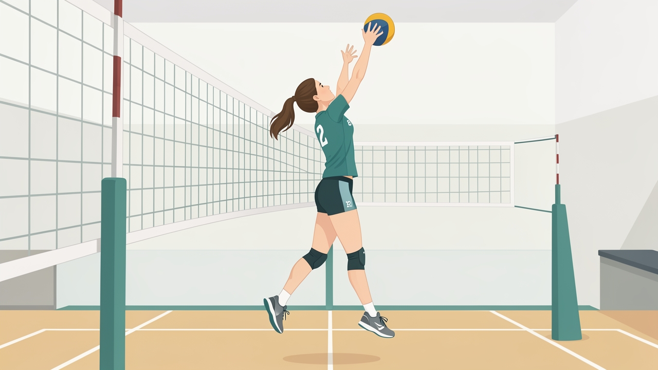
Throwers, tennis players, swimmers and volleyball players all depend on the scapulothoracic joint for both stability and mobility.

Throwers, tennis players, swimmers and volleyball players all depend on the scapulothoracic joint for both stability and mobility.

Osteology of the Scapula and Thorax

The scapula is a flat, triangular bone that serves as the attachment site for 17 muscles. Its key bony landmarks include the spine, acromion, coracoid process, glenoid fossa, medial border, lateral border, and inferior angle. The glenoid fossa is oriented slightly upward and anteriorly, an orientation that changes with scapular motion. The thorax provides the convex, mobile base upon which the scapula glides. The posterior rib cage, particularly ribs 2 through 7, forms the contact surface. The scapula’s medial border sits approximately 5–6 cm lateral to the spinous processes at rest, a distance that can vary with posture and athletic adaptation.

The Scapulothoracic Joint

The scapulothoracic joint is a physiological joint with no capsule or ligaments. Its stability is entirely dependent on muscular control and the negative pressure created between the subscapularis and the serratus anterior. The joint allows for four primary motions: elevation/depression, protraction/retraction, upward/downward rotation, and anterior/posterior tilt. In the overhead athlete, upward rotation and posterior tilt are especially critical for maintaining subacromial space and glenohumeral joint congruency.

Muscular Anatomy and Function

The scapulothoracic muscles can be classified as stabilizers, mobilizers, or prime movers. The trapezius (upper, middle, lower) works in force couples with the serratus anterior to produce upward rotation. The serratus anterior is the primary protractor and upward rotator, and is often weak or inhibited in athletes with shoulder pathology. The rhomboids and levator scapulae retract and elevate, respectively, while the pectoralis minor protracts and tilts the scapula anteriorly. The levator scapulae and rhomboids are frequently overactive in overhead athletes, contributing to scapular dyskinesis. The subclavius and omohyoid play lesser but important roles in scapular depression and stabilization.

Scapular Kinematics in Overhead Athletes

During the overhead throwing motion, the scapula must upwardly rotate approximately 40–60 degrees, posteriorly tilt 20–30 degrees, and externally rotate relative to the thorax. These motions are coordinated with glenohumeral abduction and external rotation. In the cocking phase, the scapula retracts and rotates upward. During acceleration, it protracts to maintain the glenoid under the humeral head. The deceleration phase requires eccentric control of the scapular retractors to dissipate forces. Any deviation from normal kinematics—such as excessive anterior tilt or insufficient upward rotation—can lead to impingement, labral tears, or rotator cuff pathology.

Clinical Assessment of Scapular Position and Motion

A systematic assessment begins with observation of static posture. The clinician notes scapular asymmetry, winging, or tilting. The distance from the medial border to the spine is measured in neutral and with hands on hips. Dynamic assessment includes the Scapular Assistance Test (SAT) and Scapular Retraction Test (SRT). The clinician should evaluate scapular motion during active arm elevation in the scapular plane, looking for early winging, delayed upward rotation, or excessive elevation. The Kibler classification system grades scapular dyskinesis as Type I (inferior angle prominence), Type II (medial border prominence), Type III (superior border prominence), or Type IV (symmetric motion). Overhead athletes often exhibit Type II or Type III patterns.

Measurement Techniques and Normative Values

Quantitative measures are essential for tracking progress. Scapular upward rotation can be measured with a digital inclinometer or goniometer at 0°, 60°, 90°, and 120° of arm elevation. Normative values for healthy adults show approximately 5–10° of upward rotation at rest, increasing to 30–50° at 120° of elevation. Lateral scapular slide test (LSST) measures the distance from the inferior angle to the nearest spinous process at 0°, 45°, and 90° of abduction. A side-to-side difference greater than 1.5 cm is considered abnormal. Posterior tilt can be assessed using a palpation meter or 3D motion analysis, though clinical use is limited. Scapular protraction is measured from the acromion to the wall or table. In overhead athletes, a protraction of 8–10 cm is common; asymmetry >1.5 cm warrants attention.

Common Scapular Dysfunctions in Overhead Athletes

Scapular dyskinesis is present in up to 68% of overhead athletes with shoulder pain. The most common patterns include:

  • SICK scapula (Scapular malposition, Inferior medial border prominence, Coracoid pain, and dyskinesis of scapular movement): characterized by a dropped, protracted, and anteriorly tilted scapula.
  • Scapular winging: medial or lateral winging due to serratus anterior or trapezius weakness.
  • Scapulothoracic bursitis: often secondary to repetitive friction or muscle imbalance.
  • Thoracic outlet syndrome: can be exacerbated by scapular depression and protraction.

Rehabilitation Principles

Rehabilitation of the scapulothoracic region in overhead athletes follows a progressive model: (1) restore mobility of the thoracic spine and soft tissue, (2) activate and strengthen inhibited muscles (serratus anterior, lower trapezius), (3) normalize force couples, (4) integrate into sport-specific movements. Specific protocols include:

  • Phase 1 (Mobility): Thoracic spine extension mobilization (foam roller, extension over a ball), pectoralis minor stretching, and latissimus dorsi release.
  • Phase 2 (Activation): Prone Y, T, W exercises; serratus anterior punches; wall slides; and supine scapular retraction.
  • Phase 3 (Strength and Control): Side-lying external rotation with retraction, prone horizontal abduction, and push-up plus.
  • Phase 4 (Sport-Specific): Throwing progression with emphasis on scapular retraction during cocking and protraction during follow-through.

Clinicians should monitor for substitution patterns (e.g., upper trapezius dominance) and use real-time feedback (mirror, tactile, or EMG biofeedback) to ensure proper motor control.

Conclusion

The scapulothoracic joint is the critical link between the trunk and the upper extremity in overhead athletes. A thorough understanding of its anatomy, kinematics, and assessment techniques allows the physical therapist to identify subtle dysfunctions that can compromise performance and increase injury risk. By integrating normative values, clinical tests, and evidence-based rehabilitation progressions, clinicians can effectively restore optimal scapular function and return athletes to their sport with confidence.

References

Kibler, W. B., Ludewig, P. M., McClure, P. W., Michener, L. A., Bak, K., & Sciascia, A. D. (2013). Clinical implications of scapular dyskinesis in shoulder injury: The 2013 consensus statement from the 'Scapular Summit'. British Journal of Sports Medicine, 47(14), 877–885. https://doi.org/10.1136/bjsports-2013-092425

Ludewig, P. M., & Reynolds, J. F. (2009). The association of scapular kinematics and glenohumeral joint pathologies. Journal of Orthopaedic & Sports Physical Therapy, 39(2), 90–104. https://doi.org/10.2519/jospt.2009.2808

McClure, P. W., Michener, L. A., Sennett, B. J., & Karduna, A. R. (2001). Direct 3-dimensional measurement of scapular kinematics during dynamic movements in vivo. Journal of Shoulder and Elbow Surgery, 10(3), 269–277. https://doi.org/10.1067/mse.2001.112954

Michener, L. A., McClure, P. W., & Karduna, A. R. (2003). Anatomical and biomechanical mechanisms of subacromial impingement syndrome. Clinical Biomechanics, 18(5), 369–379. https://doi.org/10.1016/S0268-0033(03)00047-0

Neumann, D. A. (2017). Kinesiology of the musculoskeletal system: Foundations for rehabilitation (3rd ed.). Elsevier.

Reinold, M. M., Escamilla, R. F., & Wilk, K. E. (2009). Current concepts in the scientific and clinical rationale behind exercises for glenohumeral and scapulothoracic musculature. Journal of Orthopaedic & Sports Physical Therapy, 39(2), 105–117. https://doi.org/10.2519/jospt.2009.2835

Uhl, T. L., Kibler, W. B., Gecewich, B., & Tripp, B. L. (2009). Evaluation of clinical assessment methods for scapular dyskinesis. Arthroscopy: The Journal of Arthroscopic & Related Surgery, 25(11), 1240–1248. https://doi.org/10.1016/j.arthro.2009.06.007

The other 11 chapters, all 144 MCQs and the flashcards are in the PhysioAI app.

Practice questions

5 sample MCQs from this course. Tap a question to see the answer.

  1. A 22-year-old collegiate baseball pitcher presents with a three-week history of diffuse right shoulder pain and a sensation of 'catching' during the late cocking phase of throwing. He denies any specific traumatic event but reports that the pain has progressively worsened, now limiting his ability to throw at full velocity. On physical examination, there is tenderness over the anterolateral shoulder and a positive apprehension test at 90 degrees of abduction and external rotation. Scapular dyskinesis is noted, with winging of the inferior angle during arm elevation.

    1. Which scapular muscle is most active during the cocking phase?

    • A. Lower trapezius
    • B. Pectoralis minor
    • C. Upper trapezius
    • D. Serratus anterior
    Show answer

    Answer: D. During the cocking phase, the serratus anterior is critical for scapular protraction and upward rotation, allowing the glenoid to maintain a stable position under the humeral head. Weakness or inhibition of this muscle contributes to scapular dyskinesis and impingement-like symptoms, as seen in overhead athletes. The other options are not the primary scapular muscle active during this specific phase.

  2. A 35-year-old recreational tennis player reports right shoulder pain and a sensation of 'catching' during serves. On observation, you note that during the first 30° of abduction, the scapula elevates excessively without adequate glenohumeral movement. Further assessment reveals that during full arm elevation, the overall motion is 180°, with a specific distribution between the glenohumeral and scapulothoracic joints. What is the normal ratio of glenohumeral to scapulothoracic motion in this rhythm?

    2. What is the normal scapulohumeral rhythm ratio during arm elevation?

    • A. 1:1 ratio
    • B. Variable depending on activity
    • C. 1:2 ratio
    • D. 2:1 ratio
    Show answer

    Answer: D. Normal scapulohumeral rhythm is a 2:1 ratio, meaning for every 2° of glenohumeral motion, there is 1° of scapulothoracic motion. This results in approximately 120° of glenohumeral and 60° of scapulothoracic contribution to reach 180° of arm elevation.

  3. A 28-year-old competitive swimmer presents with right shoulder pain during overhead strokes. Physical examination reveals no obvious weakness or instability, but the clinician suspects abnormal scapular motion. To objectively assess for scapular dyskinesis, the patient is asked to perform repeated arm elevations while holding a light weight, and the examiner observes the scapular rhythm from behind.

    3. Which of the following best describes the scapular dyskinesis test?

    • A. Observing scapular motion during 3-5 repetitions of weighted arm elevation
    • B. Palpating the scapula at rest
    • C. Measuring scapular position with a goniometer
    • D. X-ray imaging only
    Show answer

    Answer: A. The scapular dyskinesis test involves dynamic observation of scapular movement during repeated weighted arm elevation to detect abnormal motion patterns. Palpation at rest or static goniometric measurements do not assess dynamic control, and X-ray is not typically used for this functional assessment.

  4. A 45-year-old construction worker presents with right shoulder pain that began after repetitive overhead lifting. He reports a sharp, localized ache over the top of the shoulder that worsens when reaching across his body to fasten his seatbelt. On examination, palpation over the AC joint elicits tenderness, and horizontal adduction reproduces his pain. Passive accessory motion testing reveals reduced posterior glide of the clavicle on the acromion.

    4. Based on the clinical presentation, which of the following best describes the indication for joint mobilization of the acromioclavicular (AC) joint?

    • A. All shoulder conditions with associated stiffness
    • B. Acute clavicle fracture with displacement
    • C. Isolated rotator cuff tears without joint involvement
    • D. AC joint hypomobility and pain with horizontal adduction/cross-body movement
    Show answer

    Answer: D. Joint mobilization of the AC joint is specifically indicated for hypomobility and pain localized to the AC joint, especially when movements like horizontal adduction (cross-body adduction) reproduce symptoms. Options A, B, and C are incorrect because AC joint mobilization is not appropriate for all shoulder conditions, acute fractures, or isolated rotator cuff pathology without AC joint involvement.

  5. A 22-year-old college baseball pitcher presents with right shoulder pain that has progressively worsened over the past month. He reports that the pain is most intense when he is preparing to throw (late cocking) and immediately after releasing the ball (deceleration). Physical exam reveals posterior shoulder tightness and mild glenohumeral internal rotation deficit (GIRD). Which phase of the pitching motion is most associated with the highest shoulder stress?

    5. During which phase of the pitching motion is shoulder stress highest?

    • A. Wind-up
    • B. All phases equally
    • C. Follow-through only
    • D. Late cocking and deceleration phases
    Show answer

    Answer: D. The late cocking and deceleration phases generate the greatest shoulder stress due to extreme external rotation and eccentric muscle forces, respectively. These phases place the highest demand on the anterior capsule and rotator cuff, leading to common overuse injuries in pitchers.

Flashcards

  • What is the scapulothoracic articulation?

    A functional joint where the scapula glides on the posterior thoracic wall over ribs 2-7, not a true anatomical joint with a capsule.

  • What is the normal scapulohumeral rhythm ratio during humeral elevation, and what are the clinical implications of dysrhythmia?

    Normal scapulohumeral rhythm is a 2:1 ratio: for every 2° of humeral elevation, the scapula upwardly rotates 1°. This coordination maintains glenohumeral joint congruence and subacromial space. Dysrhythmia—such as early scapular elevation or reduced rotation—is common in impingement and rotator cuff tears, leading to pain and altered biomechanics.

  • According to Kibler's classification, what is Type III scapular dyskinesis?

    Type III scapular dyskinesis is characterized by excessive early scapular elevation and superior translation (shrugging) during arm elevation. It is commonly associated with upper trapezius hyperactivity and lower trapezius weakness, often seen in patients with shoulder impingement or rotator cuff pathology. Clinical identification relies on visual observation of the scapular resting position and dynamic motion, noting the superior border rising excessively in the first 30-60 degrees of elevation.

  • Define joint mobilization and describe its Maitland grading system.

    Joint mobilization is a passive, skilled technique applying oscillatory or sustained movements to joints to reduce pain and improve range of motion. The Maitland grading system uses four grades: Grade I (small amplitude at beginning of range), Grade II (large amplitude within mid-range), Grade III (large amplitude into tissue resistance), and Grade IV (small amplitude at end range). Clinical application varies: lower grades for pain modulation, higher grades for stiffness.

  • During which two phases of the throwing motion does the shoulder experience the highest biomechanical stress, and what specific injuries are associated with each?

    The late cocking phase generates peak anterior shear forces (up to 400 N) and external rotation torque (~90 N·m), predisposing the labrum to SLAP tears and the anterior capsule to microtrauma. The deceleration phase imposes high eccentric loads on the posterior rotator cuff and scapular stabilizers, often leading to internal impingement and posterior labral pathology. Both phases require balanced strength and flexibility to prevent overuse injuries.

All courses