Introduction
Vestibular rehabilitation is a specialized, evidence-based form of physical therapy designed to alleviate symptoms and functional limitations resulting from vestibular disorders. The scope of vestibular rehabilitation encompasses assessment, differential diagnosis, and intervention for individuals with peripheral or central vestibular pathology, as well as for those with non‑vestibular causes of dizziness such as cervicogenic dizziness, persistent postural‑perceptual dizziness (PPPD), or multisensory deficits. The primary goals are to reduce dizziness and vertigo, improve gaze and postural stability, enhance balance and mobility, and minimize the risk of falls. Vestibular rehabilitation is not a single protocol but a tailored program that may include habituation, gaze stabilization, balance retraining, and canalith repositioning maneuvers. Its importance in healthcare is underscored by the high prevalence of dizziness—affecting 15–35% of older adults and accounting for 4–5 million emergency department visits annually in the United States—and by its effectiveness in reducing symptom burden, improving quality of life, and decreasing fall‑related injuries. This chapter provides a foundational understanding of vestibular rehabilitation, its clinical relevance, and the basic anatomy and physiology of the vestibular system necessary for evidence‑based practice.
Importance of Vestibular Rehabilitation in Healthcare
Dizziness and imbalance are among the most common complaints encountered in primary care, neurology, otolaryngology, and emergency medicine. The economic burden is substantial: direct costs for dizziness‑related care exceed $4 billion per year in the United States, with indirect costs from lost productivity and falls adding significantly. Falls are the leading cause of fatal and non‑fatal injuries in older adults, and vestibular impairment increases fall risk by 2–3 times. Vestibular rehabilitation has been shown to reduce fall incidence by 30–50% in patients with unilateral vestibular hypofunction and by similar magnitudes in those with bilateral loss when combined with balance training.
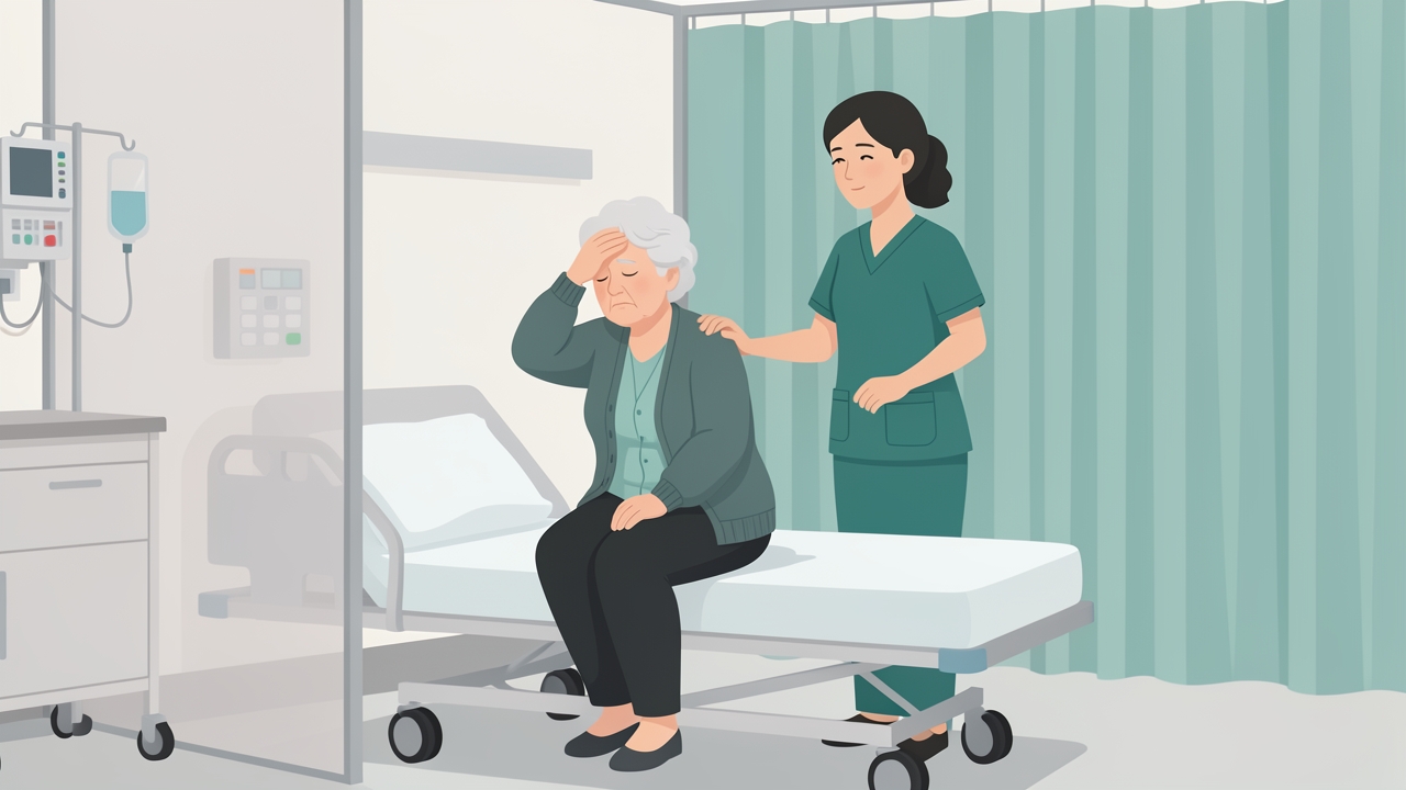
Dizziness and imbalance are among the most common complaints in emergency medicine and primary care; vestibular impairment increases fall risk 2–3 times.
Clinical outcomes are robust. For example, the Dizziness Handicap Inventory (DHI) typically improves by 15–25 points after a 4–6 week course of customized vestibular rehabilitation, with minimal clinically important difference (MCID) estimated at 11–18 points. Gaze stabilization exercises, when performed at a frequency of 3–5 times daily for 30–60 seconds per repetition, can normalize the vestibulo‑ocular reflex (VOR) gain (target >0.8) within 4–8 weeks in many patients with unilateral hypofunction. The Activities‑specific Balance Confidence (ABC) Scale shows improvements of 10–20 points, and the Timed Up and Go (TUG) test often decreases by 2–5 seconds. These metrics underscore the value of vestibular rehabilitation in restoring function and reducing disability.
Moreover, vestibular rehabilitation is cost‑effective. A 2019 systematic review found that a 6‑week program reduced healthcare utilization by 40–60% over the subsequent year, including fewer specialist visits and imaging studies. Early referral to vestibular rehabilitation is recommended by clinical practice guidelines from the American Physical Therapy Association (APTA) and the American Academy of Otolaryngology–Head and Neck Surgery, particularly for patients with acute unilateral vestibulopathy, benign paroxysmal positional vertigo (BPPV), and chronic dizziness.
Overview of the Vestibular System
The vestibular system is the sensory apparatus responsible for detecting head motion and orientation in space, thereby coordinating eye movements, postural adjustments, and spatial perception. It comprises peripheral and central components.
Peripheral Vestibular System
Semicircular Canals (SCCs): Three orthogonal canals—horizontal (lateral), anterior (superior), and posterior—detect angular acceleration. Each canal is filled with endolymph and contains a crista ampullaris with hair cells. When the head rotates, endolymph inertia deflects the cupula, altering hair cell firing. The canals are oriented at approximately 45° to the horizontal and vertical planes. The horizontal canal is most sensitive to yaw rotations, while the anterior and posterior canals respond to pitch and roll. The VOR gain for a healthy horizontal canal is typically 0.9–1.0 when measured with video head impulse testing (vHIT) at 150–200°/s peak velocity. Deflection of the cupula by even 1–2° can produce a detectable change in firing rate, which at rest is about 90–100 spikes/second.
Otolith Organs: The utricle and saccule detect linear acceleration (gravity and translation). The utricle is oriented horizontally and responds to head tilt and horizontal translation; the saccule is vertical and sensitive to vertical acceleration. Hair cells in the maculae are embedded in a gelatinous layer with otoconia (calcium carbonate crystals). Displacement of otoconia due to linear force modulates afferent firing. The threshold for otolith detection is approximately 0.1 m/s². Otolith dysfunction is assessed via cervical and ocular vestibular evoked myogenic potentials (cVEMP and oVEMP), with typical cVEMP P13‑N23 amplitudes of 50–200 μV and asymmetry ratios >30% considered abnormal.
Vestibular Nerve and Ganglia: Afferent fibers from hair cells project via the superior and inferior vestibular nerves to the vestibular ganglion (Scarpa’s ganglion) and then to the vestibular nuclei in the brainstem. Approximately 20,000 fibers innervate each labyrinth. Firing rates at rest are 50–100 spikes/s, modulated up or down with head motion.
Central Vestibular System
Vestibular Nuclei: Located in the pons and medulla, the four main nuclei (superior, lateral, medial, inferior) integrate inputs from the periphery, cerebellum, visual system, and somatosensory system. The medial vestibular nucleus is critical for VOR; the lateral nucleus (Deiters’) contributes to vestibulospinal reflexes. The cerebellum (flocculus, nodulus, uvula) fine‑tunes VOR gain and adapts to changes in vestibular function.
Vestibulo‑Ocular Reflex (VOR): The VOR stabilizes images on the retina during head movement by generating compensatory eye rotations. The three‑neuron arc (vestibular afferent → vestibular nucleus → oculomotor nucleus) has a latency of 7–10 ms. The gain (eye velocity/head velocity) should be near 1.0 for optimal vision. During head impulses at 100–200°/s, a gain <0.7 indicates hypofunction. The VOR can be tested with the head impulse test (HIT), dynamic visual acuity (DVA), and the Gaze Stabilization Test (GST). DVA loss of >2 lines on a Snellen chart suggests impairment.
Vestibulospinal Reflexes (VSR): These reflexes maintain postural stability by activating antigravity muscles. The lateral vestibulospinal tract from the lateral nucleus facilitates extensor tone; the medial vestibulospinal tract from the medial nucleus controls neck muscles. Postural sway measures (e.g., using the Modified Clinical Test of Sensory Integration and Balance, mCTSIB) quantify VSR function. Normative sway on firm surface with eyes open is <1.0 cm; with eyes closed on foam, sway may increase to 3–5 cm in healthy adults but >6 cm suggests vestibular dysfunction.
Sensory Integration: The central nervous system weights visual, somatosensory, and vestibular inputs to maintain balance. A typical sensory organization test (SOT) from computerized dynamic posturography shows equilibrium scores >90 on condition 1 (firm, eyes open) and >60 on condition 5 (foam, eyes closed). Vestibular rehabilitation aims to recalibrate these sensory weights, especially when visual or somatosensory cues are unreliable.
Clinical Decision‑Making in Vestibular Rehabilitation
Assessment begins with a thorough history (onset, duration, triggers, associated symptoms) and physical examination including orthostatic vital signs, ocular motor testing (smooth pursuit, saccades, optokinetic nystagmus), positional testing (Dix‑Hallpike, roll test), and dynamic balance.
Key Assessment Tools:
- Dizziness Handicap Inventory (DHI): 25‑item self‑report; scores 0–100; MCID 11–18 points.
- Activities‑specific Balance Confidence (ABC) Scale: 16 items; scores 0–100; MCID 8–13 points.
- Vestibular Activities and Participation Measure (VAP): 34 items; assesses participation restrictions.
- Modified Clinical Test of Sensory Integration and Balance (mCTSIB): Four conditions (firm/foam × eyes open/closed); timed up to 30 seconds; ability to hold >30 s on condition 4 (foam, eyes closed) is normal.
- Video Head Impulse Test (vHIT): Measures VOR gain for each canal; abnormal if gain <0.7 or corrective saccades present.
- Caloric Testing: Bithermal (30°C and 44°C) irrigations; normal response (slow phase velocity) 10–30°/s; unilateral weakness >25% is abnormal.
- Vestibular Evoked Myogenic Potentials (VEMP): cVEMP (sternocleidomastoid) for saccular function; oVEMP (inferior oblique) for utricular function.
Outcome Measures and Protocols:
- Gaze Stabilization Exercises: The patient moves the head horizontally or vertically at 0.5–1.0 Hz while focusing on a target. For unilateral hypofunction, perform 3–5 sets of 30–60 seconds per direction, 3–5 times daily. Progression: increase speed to 1.5–2.0 Hz, add background motion, or reduce target size.
- Habituation Exercises: For motion‑provoked dizziness, identify the specific movement that triggers symptoms (e.g., bending forward, rolling in bed). Perform the movement 2–3 times per session, 2–3 times daily, allowing symptoms to subside fully between repetitions. The goal is to reduce symptom intensity by 50% over 2–4 weeks.
- Balance Retraining: Start with static standing (feet apart, eyes open) for 30 seconds, progress to tandem stance (30 s), single‑leg stance (10–20 s), and dynamic tasks (walking with head turns, reaching). Incorporate sensory manipulation (foam, compliant surfaces, eyes closed). Parameters: 2–3 sets of 5–10 repetitions, 2–3 times daily.
- Canalith Repositioning Maneuvers (CRM): For posterior canal BPPV, the Epley maneuver is performed. Success rate >80% after one treatment; repeated up to 3 sessions if needed. The Semont maneuver is an alternative. For horizontal canal BPPV, the barbecue roll or Gufoni maneuver is used.
- Cawthorne‑Cooksey Exercises: A generic program of eye, head, and body movements performed 2–3 times daily for 5–10 minutes. While less customized, it is useful for patients with chronic, non‑localized dizziness. Progression: increase speed and complexity over 4–6 weeks.
Clinical Decision‑Making Algorithm:
- Identify the underlying diagnosis (e.g., unilateral hypofunction, BPPV, PPPD, central disorder).
- Choose the primary intervention: habituation for motion‑provoked symptoms, gaze stabilization for VOR deficits, balance training for postural instability, CRM for BPPV.
- Set frequency and duration: typically 2–3 sessions per week for 4–8 weeks, with a home exercise program performed daily.
- Monitor progress every 2–4 weeks using DHI, ABC, vHIT gain, or mCTSIB. If no improvement after 4 weeks, reassess for alternative diagnoses or adherence issues.
- Discharge criteria: DHI <20, ABC >80%, vHIT gain >0.8, mCTSIB condition 4 >30 seconds, and patient reports minimal functional limitation.
References
- Herdman SJ, Clendaniel RA. Vestibular Rehabilitation. 4th ed. Philadelphia: F.A. Davis; 2014.
- Hall CD, Herdman SJ, Whitney SL, et al. Vestibular rehabilitation for peripheral vestibular hypofunction: an evidence‑based clinical practice guideline. J Neurol Phys Ther. 2016;40(2):124‑155.
- Bhatt H, Bhatt S, Bhatt V. Vestibular rehabilitation: a review. Indian J Otol. 2012;18(3):109‑114.
- Whitney SL, Sparto PJ, Furman JM. Vestibular rehabilitation. Phys Med Rehabil Clin N Am. 2010;21(4):813‑829.
- McDonnell MN, Hillier SL. Vestibular rehabilitation for unilateral peripheral vestibular dysfunction. Cochrane Database Syst Rev. 2015;(1):CD005397.
- Strupp M, Dlugaiczyk J, Ertl‑Wagner B, et al. Vestibular disorders: diagnosis, new classification, and treatment. Dtsch Arztebl Int. 2020;117(17):300‑310.
- Cohen HS, Kimball KT. Effectiveness of treatments for benign paroxysmal positional vertigo of the posterior canal. Otol Neurotol. 2005;26(5):1033‑1040.
- Rine RM, Schubert MC, Whitney SL, et al. Vestibular rehabilitation for children and adolescents: a systematic review. Pediatr Phys Ther. 2018;30(3):190‑200.