This chapter outlines the pathophysiology, presentation, assessment and staged physiotherapy management of ischemic, hemorrhagic and transient cerebrovascular events, using clinical reasoning that applies across the stroke continuum.
Pathophysiology and Stroke Types
Stroke is a sudden focal neurological deficit of vascular origin lasting more than 24 hours, or shorter if imaging shows infarction.
- Ischemic stroke (about 85%) is caused by thrombotic, embolic (e.g., atrial fibrillation) or small-vessel lacunar occlusion. Core infarct is surrounded by a penumbra of hypoperfused but salvageable tissue, which is the rationale for early reperfusion (IV alteplase within 4.5 h, thrombectomy up to 24 h in selected patients) and for avoiding hypotension and hypoxia in the early days.
- Hemorrhagic stroke (about 15%) includes intracerebral hemorrhage (hypertensive, amyloid) and subarachnoid hemorrhage (SAH). Risks include raised intracranial pressure, rebleeding and vasospasm (days 4–14 after SAH).
- Transient ischemic attack (TIA) is a transient episode of focal dysfunction without acute infarction on imaging. It is a medical emergency because it warns of imminent stroke.
Recovery depends on spontaneous neurological recovery (greatest in the first 3 months) and experience-dependent neuroplasticity: cortical reorganisation, perilesional recruitment and reduced diaschisis, all driven by intensive, task-specific practice.
Vascular Territories and Clinical Presentation
| Territory | Typical presentation |
|---|---|
| Left MCA | Right hemiparesis (face/arm > leg), right sensory loss, aphasia (global if both Broca and Wernicke areas are involved), right hemianopia, apraxia |
| Right MCA | Left hemiparesis, left hemisensory loss, unilateral neglect, anosognosia, impaired spatial perception, left hemianopia |
| ACA | Contralateral leg > arm weakness, abulia, urinary incontinence |
| PCA | Contralateral hemianopia, memory deficits |
| Vertebrobasilar | Ataxia, vertigo, dysphagia, cranial nerve signs, crossed deficits |
| Lacunar | Pure motor or pure sensory stroke, ataxic hemiparesis |
A 60–65-year-old man with sudden left arm and leg weakness suggests a right-hemisphere lesion. Look for neglect, poor safety awareness and impulsivity, which influence fall risk and handling. A 68-year-old man with left MCA stroke has right hemiparesis plus global aphasia, so comprehension and expression are both impaired. Use demonstration, gesture, pictures and consistent routines rather than relying on verbal instruction.
Early motor signs typically follow flaccidity (hypotonia) with later spasticity and synergy patterns. Typical upper-limb patterns are scapular retraction, shoulder adduction/internal rotation, elbow flexion, forearm pronation and finger flexion. Typical lower-limb patterns are hip extension/adduction/internal rotation, knee extension and plantarflexion/inversion.
Acute Medical Considerations and Red Flags
Before treating, review imaging, blood pressure parameters, neurological stability and medications.
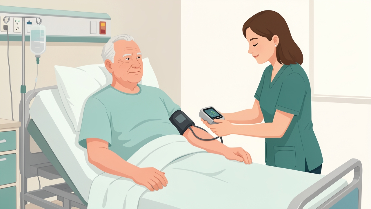
Before treating, review blood pressure, neurological stability and medications. After thrombolysis, BP is usually kept below 180/105 mmHg for 24 h.
- Red flags: declining GCS or NIHSS, new severe headache, vomiting, pupil asymmetry, seizure, chest pain, calf swelling (DVT), SpO₂ <90% (or <88% in COPD), resting HR >100–110 bpm.
- Post-thrombolysis: BP is usually kept <180/105 mmHg for 24 h. Hold mobilisation until cleared by the medical team.
- Hemorrhagic stroke: BP targets are set by the physician (often SBP <140–160 mmHg). Avoid Valsalva and sustained isometric effort. For SAH, mobilise only after the aneurysm is secured.
- Contraindications to progressing activity: uncontrolled hypertension, unstable cardiac rhythm, early neurological deterioration, raised intracranial pressure.
- Complications to screen for: aspiration (dysphagia), shoulder pain or subluxation, DVT, pressure injury, depression, falls.
Assessment: Body Structure and Function
Level of consciousness and severity
- NIHSS (0–42): 0 = no deficit, 1–4 minor, 5–15 moderate, 16–20 moderate–severe, 21–42 severe.
Motor and tone
- MRC/MMT (0–5): 3 = full range against gravity, 4 = against moderate resistance.
- Modified Ashworth Scale: 0, 1, 1+, 2, 3, 4.
- Brunnstrom stages 1–6: flaccidity → synergy → voluntary movement out of synergy.
- Chedoke-McMaster Stages of Motor Recovery (1–7) for arm, hand, leg and foot.
- Fugl-Meyer Assessment: motor total 100 (UE 66, LE 34). MCID is about 5–7 points for the UE.
- Joint range: goniometry, for example shoulder flexion ≥120° and ankle dorsiflexion to neutral, to prevent contracture.
Sensory, perceptual, cognitive and communication
- Light touch, proprioception and stereognosis, assessed by comparison with the unaffected side.
- Neglect: line bisection, star cancellation, Catherine Bergego Scale.
- Cognition: MoCA (normal ≥26/30).
- Communication: Frenchay Aphasia Screening Test or Western Aphasia Battery, and a speech-language pathology referral. A 50-year-old woman with communication difficulty needs her comprehension, expression and dysarthria or apraxia differentiated, because this determines how you instruct and educate her.
Cardiorespiratory
- Resting vital signs, Borg CR10 dyspnea scale, SpO₂ monitoring (target 88–92% in COPD unless otherwise prescribed).
Assessment: Activity and Participation
| Domain | Measure | Key reference values |
|---|---|---|
| Disability | modified Rankin Scale (0–6) | 0–2 = independent; 3–5 = dependent |
| ADL | Barthel Index (0–100) | <60 = substantial dependence; ≥90 = largely independent |
| ADL | FIM (18–126) | MCID about 22 points |
| Postural control | Postural Assessment Scale for Stroke; Trunk Impairment Scale (0–23) | Used acutely |
| Balance | Berg Balance Scale (0–56) | ≤45 = fall risk; MCID about 5–7 |
| Mobility | Timed Up and Go | >14 s = elevated fall risk after stroke |
| Gait speed | 10-Metre Walk Test | <0.4 m/s household; 0.4–0.8 limited community; >0.8 m/s community; MCID 0.10–0.16 m/s |
| Endurance | 6-Minute Walk Test | Healthy adults about 400–700 m; MCID about 34 m |
| Upper limb | Action Research Arm Test (0–57), Box and Block Test | MCID ARAT about 5.7 |
| Reintegration | Stroke Impact Scale | Participation and quality of life |
A 55-year-old woman struggling with ADLs should be assessed with the Barthel Index or FIM, then analysed component by component (transfers, dressing, toileting) to identify whether the limit is weakness, tone, neglect, apraxia or endurance.
Physiotherapy Management: Staged Approach
Hyperacute/acute (0–7 days)
- Positioning: 2-hourly changes, supine/side-lying, with the affected shoulder supported in protraction and slight external rotation. Avoid prolonged hip external rotation.
- Early mobilisation: begin within 24–48 h once medically stable, and use short, frequent sessions. The AVERT trial found that very early, high-dose mobilisation (first session <24 h, three extra sessions/day) led to less favourable 3-month outcomes than usual care.
- Progression: supported sitting → sit-to-stand → transfers → short assisted walking.
- Respiratory care: active cycle of breathing, upright positioning and early mobilisation. For a COPD patient, add pursed-lip breathing, energy conservation and rest breaks with SpO₂ monitoring.
- Other measures: passive ROM, intermittent pneumatic compression if immobile, and dysphagia precautions (nil by mouth until screened).
Early subacute (1–3 months): the window of maximal plasticity
- Task-specific, high-repetition training: sit-to-stand, stepping, reaching and grasping. Aim for 30–60 min of active practice per day, with total inpatient therapy up to about 3 h/day.
- Gait: body-weight-supported or overground training, with an AFO for foot drop. Progress speed, then add obstacles and dual tasking.
- Strengthening: 2–3 sets of 8–12 repetitions at about 60–80% of 1RM, 2–3 days/week.
- Upper limb: constraint-induced movement therapy (CIMT) for patients with ≥10° wrist extension and ≥10° finger extension (EXCITE criteria), with mirror therapy, task-oriented training and, where indicated, electrical stimulation.
- Balance: progress from sitting to standing, narrowing the base of support, perturbation training and treadmill work.
- Tone: stretching, positioning and splinting. Refer for botulinum toxin if focal spasticity limits function.
Late subacute and chronic (>3–6 months)
- Aerobic exercise: 20–60 min, 3–5 days/week, at 40–70% HRR or RPE 11–14, using a treadmill or cycle ergometer. Screen cardiac status first.
- Community reintegration: circuit class training, fall prevention, driving and return-to-work advice.
- Maintenance: continued gains are possible in the chronic phase with sufficient intensity.
Condition-Specific Considerations
- Hemorrhagic stroke with balance and mobility deficits (70-year-old woman): after medical clearance, treat as for ischemic stroke but monitor BP closely. Start balance work in supported sitting, then progress to standing with a harness or frame, then dynamic and dual-task challenges. Document the BMI, orthostatic BP response and vestibular or cerebellar signs.
- Aphasia with right hemiparesis: use demonstration, mirroring, visual schedules and one-step commands. Do not assume that "yes/no" answers are reliable. Maintain dignity and avoid childlike language.
- Right-hemisphere stroke: compensate for neglect by cueing, placing objects on the affected side and using verbal scanning strategies. Supervise closely, as impulsivity raises fall risk.
- Shoulder care: avoid overhead pulleys and traction on the hemiplegic arm. Limit passive flexion/abduction to about 90° unless the scapula upwardly rotates and the humerus is externally rotated. Assess subluxation (fingerbreadth gap), and consider a sling only for short periods during mobilisation.
Managing TIA
A 60- or 70-year-old man with a TIA and no residual deficits still requires urgent medical assessment.
- ABCD² score: Age ≥60 (1), BP ≥140/90 (1), Clinical features (unilateral weakness 2, speech disturbance 1), Duration (≥60 min 2, 10–59 min 1), Diabetes (1). Scores of 0–3, 4–5 and 6–7 are low, moderate and high risk, with 2-day stroke risk of about 1%, 4% and 8%.
- Physiotherapist's role: recognise symptoms, escalate immediately (same-day specialist referral), and screen balance, gait speed and cardiovascular fitness.
- Longer-term role: advise on secondary prevention through graded aerobic activity (at least 150 min/week moderate intensity), BP control, smoking cessation and medication adherence.
Evidence and Clinical Pearls
- Treatment intensity and task-specificity are the strongest predictors of motor gain (Veerbeek 2014).
- Early mobilisation is beneficial, but not very early, very high dose (AVERT).
- Most motor recovery occurs in the first 3 months, but plateaus are not absolute, so reassess regularly.
- Use valid measures at baseline and at standard intervals (e.g., admission, discharge, 3 months) and compare changes with MCID values rather than relying on impressions.
- Learned non-use can be prevented by enforcing use of the affected limb.
- Avoid facilitating abnormal synergies by forcing resisted effort in patients with severe tone.
- Falls: the greatest risk is in the first weeks after discharge. Prescribe home exercises and assess the home environment.
- Treat fatigue and depression as rehabilitation barriers. Screen with the PHQ-9 and refer.
- Educate caregivers early, using teach-back methods.
Summary
Stroke rehabilitation begins in the first days with safe positioning, respiratory care and early mobilisation. It progresses to high-intensity, task-specific training as the patient stabilises. Accurate classification (ischemic, hemorrhagic, TIA), identification of the vascular territory, and standardised measures (NIHSS, Fugl-Meyer, Berg, 10MWT, Barthel, mRS) allow therapists to set goals, adjust dosage and recognise red flags. Comorbidities such as COPD, and impairments such as aphasia or neglect, shape how each intervention is delivered. TIA, although it leaves no deficit, demands urgent referral and secondary prevention.
References
- Winstein CJ, Stein J, Arena R, et al. Guidelines for adult stroke rehabilitation and recovery: a guideline for healthcare professionals from the American Heart Association/American Stroke Association. Stroke. 2016;47(6):e98–e169.
- Powers WJ, Rabinstein AA, Ackerson T, et al. Guidelines for the early management of patients with acute ischemic stroke: 2019 update. Stroke. 2019;50(12):e344–e418.
- AVERT Trial Collaboration Group. Efficacy and safety of very early mobilisation within 24 h of stroke onset (AVERT): a randomised controlled trial. Lancet. 2015;386(9988):46–55.
- Hebert D, Lindsay MP, McIntyre A, et al. Canadian stroke best practice recommendations: stroke rehabilitation practice guidelines, update 2015. Int J Stroke. 2016;11(4):459–484.
- Veerbeek JM, van Wegen E, van Peppen R, et al. What is the evidence for physical therapy poststroke? A systematic review and meta-analysis. PLoS One. 2014;9(2):e87987.
- Wolf SL, Winstein CJ, Miller JP, et al. Effect of constraint-induced movement therapy on upper extremity function 3 to 9 months after stroke: the EXCITE randomized clinical trial. JAMA. 2006;296(17):2095–2104.
- Fugl-Meyer AR, Jääskö L, Leyman I, Olsson S, Steglind S. The post-stroke hemiplegic patient. 1. A method for evaluation of physical performance. Scand J Rehabil Med. 1975;7(1):13–31.
- Johnston SC, Rothwell PM, Nguyen-Huynh MN, et al. Validation and refinement of scores to predict very early stroke risk after transient ischaemic attack. Lancet. 2007;369(9558):283–292.