This chapter links the neurobiology of recovery (neuroplasticity and motor learning) to the assessment and staged management of people with stroke, unilateral neglect and multiple sclerosis, using validated outcome measures to guide each decision.
Neuroplasticity and Recovery After Stroke
Neuroplasticity is the nervous system's capacity to reorganise structure and function in response to experience or injury. After ischaemic stroke, spontaneous biological recovery is greatest in the first 3 months. It is driven by resolution of diaschisis and oedema, perilesional synaptogenesis, axonal sprouting, unmasking of latent pathways and cortical map remapping. Most motor recovery plateaus by about 3–6 months, although task-specific gains can continue beyond this.
Activity-dependent plasticity relies on long-term potentiation (LTP) and long-term depression (LTD). Repetitive, meaningful, progressively challenging practice drives use-dependent plasticity. The same principle applies outside stroke. A 58-year-old with incomplete cervical SCI (C5, AIS D) who completes 6 weeks of task-specific upper-limb training and shows increased contralateral primary motor cortex activation on fMRI is demonstrating training-induced cortical reorganisation. It is not simply spontaneous recovery.
Plasticity is also experience-dependent and competitive. Learned non-use and compensatory strategies, such as trunk substitution for reach, can reinforce maladaptive patterns, so therapists must grade tasks to encourage true recovery wherever possible.
Motor Learning Principles
Fitts and Posner describe three stages: cognitive (understanding the task, high variability, reliance on verbal cues), associative (refinement, reduced error and variability) and autonomous (automatic performance with low attentional demand). A patient 3 weeks after left MCA stroke who reaches for a cup with highly variable trajectories and needs maximal verbal cues is in the cognitive stage.
Management at this stage includes:
- Simple, clear instructions and a demonstration, with an external focus of attention (for example, "reach toward the cup" rather than "extend your elbow").
- Blocked, then serial, practice of a single task, with manual or environmental guidance reduced as accuracy improves.
- Knowledge of results (KR) and knowledge of performance (KP) given frequently at first and then faded to about 33–50% of trials, to avoid dependence on feedback.
- Progression to random, variable practice and dual-task conditions in the associative stage to promote retention and transfer.
Clinical Presentation: Stroke Syndromes and Motor Planning
Left MCA infarct typically produces right hemiparesis (face and arm more than leg), sensory loss and aphasia (expressive, receptive or global). Right MCA infarct produces left hemiparesis, left neglect, anosognosia, impaired visuospatial skills and impulsivity. Right-sided weakness with expressive aphasia localises to the left hemisphere, whatever the label on a referral. Always reconcile the deficit pattern with the lesion side.
Motor planning deficits after left-hemisphere stroke include ideomotor apraxia and difficulty initiating gait. A patient 4 weeks post-stroke who stands with minimal assistance but takes several seconds to begin stepping has impaired anticipatory postural adjustment and motor program initiation. He is not weak in the usual sense. Management uses external cueing, which bypasses impaired internally generated, basal ganglia–supplementary motor area (SMA) pathways.
In the acute flaccid stage (Brunnstrom stage 1), muscle tone is reduced, there are no voluntary movements and the glenohumeral joint is vulnerable to subluxation and traction injury. Typical recovery proceeds through emerging spasticity and synergies (stages 2–3) to selective movement (stages 4–6).
Unilateral Neglect and Postural Disorders
Neglect is failure to report, respond or orient to stimuli contralateral to the lesion, and is not explained by sensory or motor deficit. It follows right parietal, frontal or thalamic lesions and predicts poorer functional outcome and longer length of stay. A 68-year-old with right MCA stroke, left hemiparesis and reduced attention to left-sided stimuli fits this picture. He is also at high risk of falls and left-side injury.
Contraversive pushing presents with a posterior pelvic tilt and a lean toward the hemiparetic side, with active resistance to correction. This differs from simple weakness-related lean. The patient is using the non-paretic limbs to push because of a disturbed perception of body orientation (subjective postural vertical), while visual vertical is usually intact.
Multiple Sclerosis: Pathophysiology and Spasticity
MS is an immune-mediated demyelinating and neurodegenerative disease. Conduction block in demyelinated axons causes fatigue, weakness and spasticity. Spasticity is a velocity-dependent increase in tonic stretch reflexes (clasp-knife) due to loss of descending inhibition. This differs from rigidity, which is constant, non-velocity-dependent and present in both directions.
A woman with 10 years of relapsing-remitting MS and 2 days of increased leg stiffness (Modified Ashworth Scale [MAS] 3 in right quadriceps and plantarflexors) needs a cause identified before any exercise prescription. The usual triggers are urinary tract infection, constipation, pressure injury, ill-fitting orthosis, heat (Uhthoff phenomenon) and fever. A relapse is defined as new or worsening neurological symptoms lasting more than 24 hours, occurring at least 30 days after a previous attack, in the absence of fever or infection. A pseudo-relapse is a transient worsening of symptoms caused by infection or heat that resolves when the trigger is treated.
Assessment and Outcome Measures
Impairment
- Fugl-Meyer Assessment (upper extremity 0–66, lower extremity 0–34). The minimal clinically important difference (MCID) for the upper extremity is about 5 points.
- MAS (0, 1, 1+, 2, 3, 4). A score of 3 means considerable increase in tone with passive movement difficult.
- Tardieu Scale: R1 (catch angle at fast velocity) and R2 (full passive range at slow velocity). A large R2–R1 difference indicates a dynamic, reflex component. Normal ankle dorsiflexion with the knee extended is about 20°.
- NIHSS (0–42) for stroke severity. The SAFE score (shoulder abduction and finger extension, each graded 0–5 on MRC) at 72 hours predicts upper-limb recovery.
Activity and participation
- Berg Balance Scale (0–56). A score below 45 indicates increased fall risk.
- 10-Metre Walk Test. Healthy comfortable speed is about 1.2–1.4 m/s. Perry's classification is below 0.4 m/s for household ambulation, 0.4–0.8 m/s for limited community and above 0.8 m/s for community ambulation. A change of 0.10 m/s is a small meaningful change and 0.20 m/s is substantial.
- 6-Minute Walk Test. The MCID after stroke is about 34 m.
- Timed Up and Go. More than 14 seconds suggests falls risk after stroke.
- Action Research Arm Test (0–57) and Box and Block Test (healthy adults approximately 75–85 blocks/min).
- Barthel Index (0–100), modified Rankin Scale (0–6) and FIM.
- For MS: EDSS, Timed 25-Foot Walk, 9-Hole Peg Test, MSWS-12 and the Modified Fatigue Impact Scale.
Neglect and pushing
- Star Cancellation (maximum 54; a score of 51 or below suggests neglect).
- Line Bisection, BIT Conventional (cut-off 129/146) and the functional Catherine Bergego Scale (0–30).
- Scale for Contraversive Pushing (SCP). Pushing is present when each of the three subscales (posture, abduction/extension, resistance) scores above 0.
- Screen cognition with the MoCA (below 26 abnormal) and communication separately, so aphasia does not invalidate other testing.
Physiotherapy Management: Acute Phase (Days 0–14)
For a patient 3 days post-stroke who is medically stable and able to follow simple commands, treatment priorities are prevention of complications and graded early activity.
- Mobilisation: Begin after the first 24 hours, using short, frequent sessions. The AVERT trial showed that very early, high-dose mobilisation (within 24 hours) reduced the odds of a favourable outcome at 3 months. Monitor blood pressure and symptoms, and stop for new neurological signs, symptomatic hypotension or uncontrolled blood pressure.
- Flaccid upper limb: Support the arm in positioning, with a trough, pillow or table in sitting. Do not pull on the arm during transfers or use overhead pulleys, because both increase subluxation and pain risk. Add neuromuscular electrical stimulation (NMES) to supraspinatus and posterior deltoid (for example, 20–30 minutes daily at a comfortable tetanic contraction) as an adjunct.
- Communication: Use short commands, gestures and demonstration. Coordinate with speech pathology and avoid treating non-response as non-compliance.
- Respiratory and DVT prevention: Provide positioning, early sitting and standing, and mechanical prophylaxis as ordered.
Physiotherapy Management: Subacute Phase and Specific Problems
Task-specific training. Use high-repetition, goal-directed practice, such as 30–60 minutes per session, at least 5 days per week. Examples are reach-to-grasp, sit-to-stand (aim for 3 sets of 10), step-ups and treadmill training with or without body-weight support. Constraint-induced movement therapy (CIMT) suits patients with at least 20° wrist and 10° finger extension. The EXCITE protocol used 90% waking-hour restraint with 6 hours of training for 10 weekdays. Modified CIMT uses 2–3 hours of supervised practice daily.
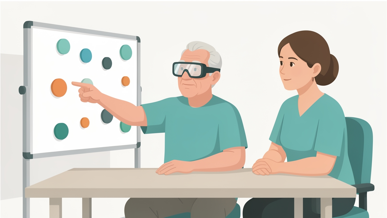
Prism adaptation for neglect: 10° rightward-shifting goggles, about 90 pointing movements per session, twice daily for 2 weeks, as an adjunct to functional training.
Gait initiation difficulty. Use rhythmic auditory cueing (metronome at 100–110% of baseline cadence), floor markers and verbal "ready-step" cues. Practise weight-shifting before stepping, and add dual-task training progressively. Fit a lightweight ankle-foot orthosis if dorsiflexor weakness causes toe drag.
Pushing. Provide visual feedback from a mirror or vertical reference, and guide the patient to actively align his midline upright. Train weight-shift toward the non-paretic side so the patient discovers an upright position, and progress to standing and gait. In the wheelchair, correct pelvic position to neutral with a firm seat and lateral supports. Do not forcibly correct the trunk against active resistance.
Neglect. Use visual scanning training, with cueing at the left margin; limb activation; trunk rotation; and environmental adaptation (place items in the right visual field initially, then progressively left). Prism adaptation uses 10° rightward-shifting goggles, about 90 pointing movements per session, twice daily for 2 weeks. The evidence for prism adaptation is inconsistent, so use it as an adjunct and not as a replacement for functional training. Ensure left-side protection during transfers.
MS spasticity. After excluding and treating triggers, use daily stretching held for 30–60 seconds, repeated 3–5 times. Add positioning, standing programs and cooling strategies. Prescribe aerobic exercise (30 minutes, twice weekly) and resistance exercise (2 sets of 10–15 for major muscle groups, twice weekly) at moderate intensity, following MS physical activity guidelines. Schedule sessions for cooler parts of the day, and use rest breaks and energy conservation. Refer to a physician for pharmacological options such as baclofen, tizanidine or botulinum toxin. Refer urgently if there is a rapid decline, new bladder or bowel dysfunction, or a sensory level.
Evidence and Clinical Pearls
- Recovery is time-sensitive. The first 3 months are the highest-yield window for intensive practice, but training should not stop at 6 months.
- Intensity and specificity matter more than modality. Doing more repetitions of the actual task is the single most consistent finding across stroke rehabilitation guidelines.
- Predict, then plan. A SAFE score of 5 or more at 72 hours with intact corticospinal tract function (assessed with the PREP2 algorithm) predicts good upper-limb recovery, whereas poor motor evoked potential response predicts a limited outcome and favours compensatory strategies.
- Use the ICF to frame goals across body function, activity and participation, and to choose outcome measures at each level.
- Stretching alone does not produce durable change in spasticity. Combine it with active task practice.
- Neglect, apraxia and pushing are cognitive-perceptual problems. Treating them as weakness reduces effectiveness.
- A sudden change in tone in MS is infection until proven otherwise.
Summary
Recovery after stroke and in other neurological conditions rests on use-dependent plasticity, which is shaped by motor learning stage, practice structure and feedback. Accurate lesion localisation, validated measures (Fugl-Meyer, MAS, Berg, 10MWT, SCP, Star Cancellation, EDSS) and staged management protect the patient early, then build function through high-dose task-specific training. Neglect, pushing, motor planning deficits and MS spasticity each require targeted strategies and attention to red flags.
References
- Winstein CJ, Stein J, Arena R, et al. Guidelines for adult stroke rehabilitation and recovery: a guideline for healthcare professionals from the American Heart Association/American Stroke Association. Stroke. 2016;47(6):e98–e169.
- AVERT Trial Collaboration Group. Efficacy and safety of very early mobilisation within 24 h of stroke onset (AVERT): a randomised controlled trial. Lancet. 2015;386(9988):46–55.
- Wolf SL, Winstein CJ, Miller JP, et al. Effect of constraint-induced movement therapy on upper extremity function 3 to 9 months after stroke: the EXCITE randomized clinical trial. JAMA. 2006;296(17):2095–2104.
- Schmidt RA, Lee TD, Winstein C, Wulf G, Zelaznik HN. Motor Control and Learning: A Behavioral Emphasis. 6th ed. Human Kinetics; 2018.
- Kwakkel G, Lannin NA, Borschmann K, et al. Standardized measurement of sensorimotor recovery in stroke trials: consensus-based core recommendations from the Stroke Recovery and Rehabilitation Roundtable. Int J Stroke. 2017;12(5):451–461.
- Stinear CM, Byblow WD, Ackerley SJ, Smith MC, Borges VM, Barber PA. PREP2: a biomarker-based algorithm for predicting upper limb function after stroke. Ann Clin Transl Neurol. 2017;4(11):811–820.
- Latimer-Cheung AE, Pilutti LA, Hicks AL, et al. Effects of exercise training on fitness, mobility, fatigue, and health-related quality of life among adults with multiple sclerosis: a systematic review to inform guideline development. Arch Phys Med Rehabil. 2013;94(9):1800–1828.
- Rossetti Y, Rode G, Pisella L, et al. Prism adaptation to a rightward optical deviation rehabilitates left hemispatial neglect. Nature. 1998;395(6698):166–169.