Learning Outcomes
By the end of this chapter, you should be able to:
- Describe the mechanical and neural determinants of ventilation at rest and during exercise.
- Explain how ventilation-perfusion matching and diffusion govern arterial oxygenation.
- Identify the principal mechanisms that limit exercise in chronic respiratory disease.
- Select and interpret spirometry, gas exchange, and exercise tests used in pulmonary rehabilitation.
- Apply screening criteria and a staged exercise prescription with defined dosage parameters.
Respiratory Anatomy and Mechanics
The respiratory system moves air between the atmosphere and the alveoli, where gas exchange occurs across a membrane of roughly 0.5 µm thickness. Its pump consists of the diaphragm (the primary inspiratory muscle, innervated by C3-C5 via the phrenic nerve), the external intercostals, and the accessory muscles of inspiration: scalenes, sternocleidomastoid, and serratus anterior. Expiration is largely passive at rest but becomes active during exercise, when the abdominal muscles and internal intercostals contribute.
Lung recoil and chest wall compliance determine the work of breathing. In emphysema, loss of elastic recoil increases static compliance and produces dynamic hyperinflation: expiratory flow limitation prevents complete exhalation, so end-expiratory lung volume (EELV) rises during exercise and shifts the tidal breath toward the flatter, less efficient part of the pressure-volume curve. The inspiratory muscles then operate at a mechanical disadvantage. In restrictive disease, reduced compliance raises the elastic work of breathing, while in neuromuscular disease the pump itself is weakened.
Ventilation and Gas Exchange at Rest and During Exercise
Minute ventilation (V̇E) equals tidal volume multiplied by respiratory rate. Dead space (V̇D/VT, normally 0.25-0.35 at rest) is wasted ventilation that does not participate in gas exchange. During incremental exercise, V̇E rises approximately linearly with carbon dioxide production (V̇CO2) until the ventilatory threshold, after which V̇E rises disproportionately.
Oxygen uptake (V̇O2) is delivered through the cardiovascular system, but the lungs must supply oxygen and remove carbon dioxide. Normal arterial values at rest are PaO2 80-100 mmHg and PaCO2 35-45 mmHg. Ventilation-perfusion (V̇/Q) mismatch, the most common cause of hypoxaemia in lung disease, reduces PaO2 by shunting or by dead-space ventilation. During exercise, the pulmonary capillary transit time shortens (from about 0.75 s to under 0.3 s in healthy individuals), so impaired diffusion or V̇/Q mismatch can cause exercise-induced desaturation even when resting saturation is acceptable.
Pathophysiology of Exercise Limitation in Chronic Lung Disease
Exercise intolerance in chronic obstructive pulmonary disease (COPD), interstitial lung disease, and related conditions arises from several interacting mechanisms:
- Ventilatory limitation: Expiratory flow limitation and dynamic hyperinflation reduce the ventilatory reserve. Breathing reserve (BR = [maximum voluntary ventilation − peak V̇E] / MVV × 100) falls below 15% in many patients, indicating that the ventilatory system is near its ceiling.
- Gas exchange abnormality: Exercise-induced hypoxaemia occurs from V̇/Q mismatch and diffusion limitation, particularly in emphysema and interstitial disease.
- Peripheral muscle dysfunction: Deconditioning, systemic inflammation, oxidative stress, and corticosteroid exposure reduce oxidative capacity and muscle mass, so the same absolute workload demands a higher proportion of maximal effort.
- Cardiovascular involvement: Pulmonary hypertension and right ventricular dysfunction may limit cardiac output, reducing oxygen delivery.
- Symptom-driven behaviour: Dyspnoea and anxiety drive avoidance, which further reduces conditioning and creates a self-perpetuating cycle.
Clinical Presentation
Patients typically report exertional dyspnoea, reduced walking distance, fatigue, and activity avoidance. Common observations include pursed-lip breathing, use of accessory muscles, forward-leaning posture, prolonged expiration, and reduced chest expansion. Clinicians should also record weight, nutritional status (BMI below 21 kg/m² in COPD is associated with poorer prognosis), and comorbidities such as ischaemic heart disease, diabetes, and anaemia, all of which influence exercise tolerance and risk.

Common observations in chronic lung disease include pursed-lip breathing, accessory muscle use, a forward-leaning posture and prolonged expiration.
Assessment: Lung Function and Gas Exchange
Spirometry (ATS/ERS standards) provides FEV1, FVC, and FEV1/FVC ratio. Airflow obstruction is confirmed by a post-bronchodilator FEV1/FVC below 0.70 or below the lower limit of normal. Severity is graded by FEV1 % predicted: GOLD 1 ≥80%, GOLD 2 50-79%, GOLD 3 30-49%, GOLD 4 <30%.
Lung volumes and diffusion: Body plethysmography measures total lung capacity and residual volume. Transfer factor for carbon monoxide (DLCO) below 60% predicted suggests significant diffusion impairment or vascular disease.
Respiratory muscle strength: Maximal inspiratory pressure (MIP) and maximal expiratory pressure (MEP) are measured with a manometer. Values of MIP more negative than −60 cmH2O (women) and −80 cmH2O (men) are typically considered adequate; values below about 30% predicted indicate significant weakness.
Arterial blood gases: Resting PaO2 below 60 mmHg or PaCO2 above 50 mmHg indicates the need for oxygen assessment and specialist review.
Assessment: Exercise Capacity and Symptoms
Six-minute walk test (6MWT): Performed along a 30 m corridor following ATS guidelines, with SpO2, heart rate, and Borg dyspnoea ratings recorded before and after. Reference equations (Enright and Sherrill, 1998) estimate predicted distance from age, sex, height, and weight. A change of 30-50 m is generally regarded as clinically meaningful in COPD.
Incremental shuttle walk test (ISWT) and endurance shuttle walk test (ESWT): The ISWT uses an audio-paced protocol with increments each minute; the ESWT, performed at 85% of ISWT peak speed, measures endurance and is sensitive to change after rehabilitation.
Cardiopulmonary exercise testing (CPET): Provides peak V̇O2, anaerobic threshold, V̇E/V̇CO2 slope, breathing reserve, and exercise SpO2. Peak V̇O2 below 50% predicted indicates severe impairment. A V̇E/V̇CO2 slope above 35 suggests ventilatory inefficiency.
Symptom and quality-of-life measures: The Modified Medical Research Council (mMRC) dyspnoea scale (grade 0-4), the COPD Assessment Test (CAT; minimal important difference approximately 2 points), and the St George's Respiratory Questionnaire (SGRQ) are standard. Sit-to-stand tests (for example, the 1-minute sit-to-stand) provide a practical measure of lower-limb endurance in the clinic.
Physiotherapy Management: Pre-Training Screening and Safety
Before exercise, screen for contraindications and red flags:
- Absolute contraindications: Unstable angina, uncontrolled arrhythmia, severe symptomatic aortic stenosis, resting SBP above 200 mmHg or DBP above 110 mmHg, acute systemic illness, untreated pneumothorax, and recent haemoptysis without medical review.
- Red flags to stop exercise: SpO2 below 88% despite supplemental oxygen, new chest pain, presyncope or syncope, ST-segment depression of more than 1 mm, sustained heart rate above 85% of age-predicted maximum (220 − age), severe dyspnoea (Borg above 7-8 on the 0-10 scale), or new neurological signs.
- Oxygen: Patients with exercise desaturation should train with titrated oxygen to maintain SpO2 at or above 90%, using a documented prescription.
Monitor SpO2 and heart rate continuously during initial sessions, and use the Borg CR-10 scale for dyspnoea and leg effort.
Physiotherapy Management: Staged Exercise Prescription
A graded programme should progress from low-intensity conditioning to high-intensity training over an initial period of 6-12 weeks, with a maintenance component thereafter.
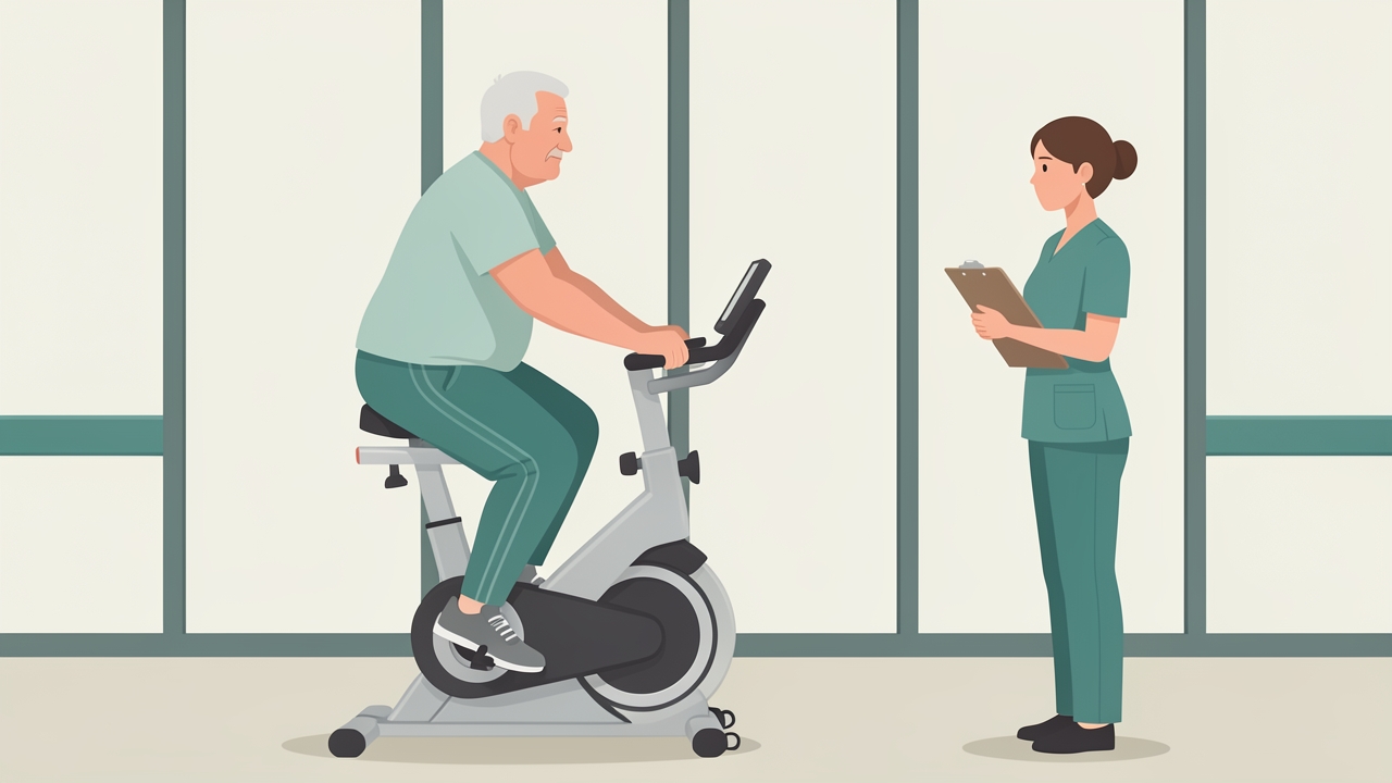
Stage 1 (weeks 1–2): 2–3 supervised sessions a week of cycle ergometry or treadmill walking, 10–15 minutes at Borg dyspnoea 2–3.
Stage 1 (weeks 1-2): Familiarisation and tolerance
- Frequency: 2-3 sessions per week, supervised.
- Mode: Cycle ergometry or treadmill walking, with upper-limb and lower-limb circuits.
- Intensity: Borg dyspnoea 2-3 (on a 0-10 scale), or 60% of the 6MWT peak heart rate response.
- Duration: 10-15 minutes continuous, progressing in five-minute blocks.
- Breathing: Pursed-lip breathing and controlled diaphragmatic breathing practised during activity.
Stage 2 (weeks 3-6): Endurance building
- Frequency: 3 sessions per week.
- Intensity: 60-70% of peak work rate (from incremental test) or 70-80% of peak heart rate, Borg 3-4.
- Duration: 20-30 minutes continuous, or interval bouts (for example, 2 minutes at 80% followed by 1 minute at 40%).
- Progression: Add 5-10 W on the cycle, or increase speed and gradient on the treadmill, when the patient completes the session without exceeding Borg 4 for the full duration.
Stage 3 (weeks 6-12): High-intensity and functional training
- Intensity: Up to 80% of peak work rate, or Borg 4-6, with high-intensity interval training used cautiously in patients with stable cardiovascular status.
- Duration: 30-45 minutes including warm-up and cool-down.
- Include strength training: 2-3 sets of 8-12 repetitions at 60-80% of one-repetition maximum for quadriceps and upper limb, or using a 10-15 repetition maximum with Borg 5-6 (dyspnoea-limited loads are acceptable).
- Include balance and functional tasks: sit-to-stand, stair climbing, and carrying tasks.
Maintenance (after week 12): At least 2 sessions per week for life, or a structured home programme with regular telephone or telehealth review, to preserve gains.
Reassess the 6MWT, ISWT or ESWT, and symptom measures at completion and at 6-12 months.
Evidence and Clinical Pearls
Pulmonary rehabilitation is one of the most robustly supported interventions in chronic respiratory disease. It reduces dyspnoea and fatigue, improves exercise capacity and health status, and reduces hospital readmission after exacerbation (Puhan et al., 2016). The ATS/ERS statement (Spruit et al., 2013) recommends a minimum of 6 weeks of supervised training, with endurance and strength components.
Clinical pearls:
- Test before you prescribe. Use the 6MWT or ISWT to set training intensity; do not rely on predicted values alone.
- Dyspnoea is not harm. Breathlessness at Borg 3-5 is expected during training; sustained chest pain, desaturation, or dizziness is not.
- Assess muscle, not just lungs. Quadriceps weakness is often a stronger predictor of disability than FEV1.
- Prevent the drop-off. Adherence falls after supervised completion; plan the maintenance phase from the first session.
- Use the MCID. Interpret change scores against the minimal clinically important difference, not statistical significance alone.
Summary
Effective exercise prescription in pulmonary rehabilitation depends on understanding how the respiratory pump, gas exchange, and peripheral muscles interact to limit exercise. Assessment should combine spirometry, diffusion, respiratory muscle strength, and field or laboratory exercise tests with symptom measures. Screening must identify contraindications and red flags before training begins. A staged programme, progressing from familiarisation through endurance and strength training to maintenance, with intensity guided by Borg scores, heart rate, and oxygen saturation, provides a safe and evidence-based framework for the remainder of this course.
References
- Enright PL, Sherrill DL. (1998). Reference equations for the six-minute walk in healthy adults. American Journal of Respiratory and Critical Care Medicine, 158(5), 1384-1387.
- Holland AE, Spruit MA, Troosters T, et al. (2014). An official European Respiratory Society/American Thoracic Society technical standard: field walking tests in chronic respiratory disease. European Respiratory Journal, 44(6), 1428-1446.
- Miller MR, Hankinson J, Brusasco V, et al. (2005). Standardisation of spirometry. European Respiratory Journal, 26(2), 319-338.
- Puhan MA, Gimeno-Santos E, Cates CJ, Troosters T. (2016). Pulmonary rehabilitation following exacerbations of chronic obstructive pulmonary disease. Cochrane Database of Systematic Reviews, (12), CD005305.
- Spruit MA, Singh SJ, Garvey C, et al. (2013). An official American Thoracic Society/European Respiratory Society statement: key concepts and advances in pulmonary rehabilitation. American Journal of Respiratory and Critical Care Medicine, 188(8), e13-e64.
- Weisman IM, Zeballos RJ. (2003). ATS/ACCP statement on cardiopulmonary exercise testing. American Journal of Respiratory and Critical Care Medicine, 167(2), 211-277.
- Global Initiative for Chronic Obstructive Lung Disease (GOLD). (2024). Global Strategy for the Diagnosis, Management, and Prevention of COPD. Available at: goldcopd.org.