Neuroanatomy of Locomotor Control
Walking depends on a layered system. The cerebral cortex, particularly the supplementary motor area, premotor cortex, and primary motor cortex, plans and modulates gait, especially during obstacle negotiation, turning, and dual-task walking. The corticospinal tract descends through the posterior limb of the internal capsule and provides the direct cortical drive to distal lower-limb muscles, notably tibialis anterior. The corticoreticular and reticulospinal pathways provide bilateral, less fine-grained control of proximal and postural muscles and are important for trunk stability and synergistic movement. The cerebellum integrates proprioceptive and vestibular input to correct timing and balance, and the basal ganglia contribute to initiation and rhythm.
Below the cortex, the spinal central pattern generators (CPGs) produce the basic alternating rhythm of stepping. These networks are strongly shaped by afferent feedback from muscle spindles, Golgi tendon organs, and cutaneous receptors, which signal load and phase transitions. Successful gait after stroke therefore depends on cortical drive, intact descending pathways, and accurate sensory feedback.
Pathophysiology of Post-Stroke Gait Impairment
Most ischaemic strokes affecting ambulation involve the middle cerebral artery (MCA) territory, which supplies lateral frontal, parietal, and temporal cortex and the corona radiata. Lacunar infarcts in the posterior limb of the internal capsule are a common cause of pure motor hemiparesis. Anterior cerebral artery involvement may produce predominantly leg weakness with less arm involvement, which is relevant when planning gait-focused treatment.
The primary deficits are:
- Loss of corticospinal drive, causing weakness that is most marked in ankle dorsiflexors and hip abductors.
- Altered muscle tone, with spasticity typically developing over days to weeks through upregulated spinal reflex excitability.
- Abnormal synergies, in which the lower limb tends toward an extensor synergy (hip extension, knee extension, ankle plantarflexion, inversion).
- Impaired sensation and proprioception, which reduces the accuracy of weight-bearing and limb-position awareness.
- Reduced plantarflexor power at push-off, which is the main determinant of paretic propulsion and walking speed.
- Cardiorespiratory deconditioning, which compounds the energy cost of walking.
Neuroplasticity and the Recovery Window
Recovery is greatest in the first 3 months, with most gains in motor function occurring within the first 4 to 6 weeks, but meaningful improvement continues beyond this. Experience-dependent plasticity follows principles described by Kleim and Jones (2008): training must be specific to the desired task, repetitive, intensive, and salient to drive durable synaptic and cortical reorganisation. Recovery can involve perilesional and ipsilesional reorganisation, and in some patients contralesional structures contribute, though maladaptive patterns can also emerge. This is the rationale for early, high-repetition, task-specific gait practice rather than compensatory training alone.
Clinical Presentation of the Hemiparetic Gait
The classic hemiparetic gait includes:
- Reduced paretic stance time and early weight transfer to the non-paretic limb.
- Circumduction or hip hiking to clear the foot during swing due to inadequate dorsiflexion.
- Foot drop in swing, often with toe contact or scuffing.
- Reduced paretic propulsion, with diminished ankle push-off.
- Trunk lean toward the non-paretic side and asymmetrical arm swing.
- Reduced cadence and step length, with a shortened paretic step.
- Stance instability at the knee, such as hyperextension or genu recurvatum when quadriceps control is poor.
Compensatory strategies are common, and some (such as hip hiking or vaulting) become habitual. Identifying which deficits are impairment-driven versus compensatory informs treatment selection.
Clinical Assessment
A structured assessment should combine impairment, activity, and participation measures.
Impairment measures
- Fugl-Meyer Assessment, Lower Extremity (FMA-LE): Maximum score 34. It is widely used and responsive in stroke.
- Modified Ashworth Scale (MAS): Grades spasticity from 0 to 4, with 1+ commonly used as a threshold for clinical relevance.
- Manual muscle testing (MRC scale): Grade 3 or higher for ankle dorsiflexion and plantarflexion, hip abduction, and knee extension is generally required for functional swing clearance and stance stability.
- Ankle dorsiflexion range with knee extended and flexed, to distinguish gastrocnemius from soleus contracture.
- Sensation and proprioception: Monofilament testing and joint-position sense at the ankle and hallux.
Activity measures
- Functional Ambulation Category (FAC): 0 (non-functional) to 5 (independent on level and uneven surfaces).
- 10-Metre Walk Test (10MWT): Measured at comfortable and fast speeds. Speed categories are household ambulation below 0.4 m/s, limited community ambulation 0.4 to 0.8 m/s, and community ambulation above 0.8 m/s (Bowden et al., 2008).
- 6-Minute Walk Test (6MWT): Assesses endurance; reduced distance is a strong predictor of community mobility.
- Timed Up and Go (TUG): Times of 13.5 seconds or longer are associated with increased fall risk after stroke.
- Berg Balance Scale (BBS): Scores below 45 out of 56 indicate increased fall risk.
Gait analysis Observational gait analysis using a standard framework (for example, Perry and Burnfield, 2010) should record stance and swing phase timing, paretic step length, cadence, and trunk alignment. Video recording is useful for review. Where available, instrumented analysis (pressure mats, inertial sensors) quantifies symmetry.
Normative Values and Interpretation
Healthy adults typically walk at a self-selected speed of 1.2 to 1.4 m/s, with cadence of approximately 100 to 120 steps per minute. Post-stroke self-selected speed is commonly 0.4 to 0.8 m/s in the subacute period, with many patients plateauing below 1.0 m/s. Fritz and Lusardi (2009) describe gait speed as a "sixth vital sign," supporting its use as a single prognostic indicator. Interpret results against the patient's pre-morbid function and goals, and track change using the minimal clinically important difference (MCID), which for 10MWT in stroke is approximately 0.1 to 0.16 m/s.
Physiotherapy Management: Staged Protocol
Treatment should progress through stages guided by FAC, medical stability, and tolerance. The following timeframes are indicative.
Stage 1: Acute (days 1 to 7 post-onset, medically stable)
- Sessions of 20 to 30 minutes, once or twice daily as tolerated.
- Bed mobility, sitting balance, sit-to-stand with assistance, and standing weight-shifting.
- Early gait initiation with parallel bars or body-weight support, emphasising symmetrical weight-bearing and paretic stance.
- Positioning and range-of-motion work for the ankle, with dorsiflexion stretching if contracture risk is present.
- Introduce orthoses (ankle-foot orthosis) where foot drop prevents safe walking.
Stage 2: Subacute (approximately weeks 2 to 12, FAC 1 to 3)
- Sessions of 30 to 45 minutes, 5 days per week.
- Body-weight-supported treadmill training (BWSTT) at progressively higher speeds, starting around 0.3 to 0.5 m/s and advancing as the patient allows, with aim for high step counts.
- Targeted strengthening: resisted ankle dorsiflexion and plantarflexion, hip abduction, and knee extension, using 2 to 3 sets of 10 to 15 repetitions at moderate resistance.
- Task-specific practice: sit-to-stand, stepping over low obstacles, and turning.
- Cardiovascular conditioning at moderate intensity (commonly 40 to 60% heart rate reserve), progressing as tolerated.
Stage 3: Community transition (approximately week 12 onward, FAC 3 to 5)
- Sessions of 45 to 60 minutes, 3 to 5 days per week.
- Overground walking progressing in speed, distance, and surface (ramps, kerbs, grass).
- Stair training with a rail, progressing to a single rail or no rail.
- Dual-task training (walking while carrying, talking, or counting) and reactive balance exercises.
- Power training for plantarflexors, such as heel raises with load, to improve push-off.
- Community-based goals, including crossing roads within timed limits and navigating shops or public transport.
- Home exercise programme and referral to community exercise or stroke groups.
Dosage should emphasise high repetition and task specificity across all stages, with progression based on measured change rather than fixed timeframes.
Red Flags and Contraindications
Stop or modify treatment and refer urgently if any of the following occur:
- New or worsening neurological signs, including headache, drowsiness, new weakness, or altered speech, which may indicate haemorrhagic transformation, recurrent stroke, or hydrocephalus.
- Chest pain, palpitations, or dyspnoea out of proportion to effort.
- Resting systolic BP above 200 mmHg or diastolic above 110 mmHg, or symptomatic hypotension (drop of 20 mmHg or more systolic on standing, or dizziness).
- Unilateral calf pain, swelling, or warmth, which may indicate deep vein thrombosis.
- Unexplained fever or signs of infection such as chest changes or wound problems.
- Sudden loss of balance with new falls, which may signal a new lesion.
Relative contraindications to high-intensity training include uncontrolled arrhythmia, recent myocardial infarction, unstable angina, and significant hypoglycaemia. Monitor perceived exertion (Borg RPE 11 to 14) and heart rate throughout.
Evidence Base
Current guidance (Winstein et al., 2016) recommends that people who can walk after stroke receive gait training that is task-specific, repetitive, and of sufficient dosage, and that treadmill training with or without body-weight support can improve walking speed and endurance. Walking practice improves speed and endurance more than balance-only interventions in many trials. Lord et al. (2004) found that community ambulation was associated with factors including balance, lower limb function, and walking speed, supporting the staged emphasis on community-relevant practice. Consistent evidence supports early mobilisation, though the optimal intensity and dose remain under investigation.
Clinical Pearls
- Treat the ankle as the key joint. Dorsiflexion control during swing and plantarflexion power at push-off drive most of the improvement in paretic gait.
- Avoid training compensations. Reduce hip hiking and vaulting with cueing, mirrors, and reduced-speed practice before progressing speed.
- Use speed as a dose driver. Faster treadmill speeds (within safe limits) increase step symmetry and cardiovascular load, but the patient must be able to maintain form.
- Reassess formally every 2 to 4 weeks using the 10MWT and FAC to guide progression.
- Consider fatigue. Schedule demanding walking after rest, and monitor for post-exertional deterioration in gait quality.
- Prioritise patient goals. Community tasks such as shopping or crossing roads should drive late-stage training.
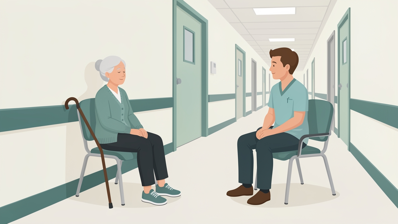
Consider fatigue: schedule demanding walking after rest, and watch for post-exertional deterioration in gait quality.
Summary
Post-stroke gait impairment reflects damage to cortical and descending motor pathways, altered spinal and sensory feedback, and secondary deconditioning. The hemiparetic gait is characterised by reduced paretic stance, foot drop, poor push-off, and compensatory patterns. Assessment should combine impairment measures (FMA-LE, MAS, MRC grading), activity measures (FAC, 10MWT, 6MWT, TUG, BBS), and observational gait analysis. Management follows a staged approach from acute mobilisation through subacute BWSTT and strengthening to community-level task training, with dosage guided by measured progress. Vigilance for red flags and appropriate contraindication screening are essential at every stage.
References
- Bowden, M. G., Balasubramanian, C. K., Behrman, A. L., & Kautz, S. A. (2008). Validation of a speed-based classification system using quantitative measures of walking performance poststroke. Neurorehabilitation and Neural Repair, 22(6), 672–675.
- Fritz, S., & Lusardi, M. (2009). White paper: "Walking speed: the sixth vital sign." Journal of Geriatric Physical Therapy, 32(2), 46–49.
- Kleim, J. A., & Jones, T. A. (2008). Principles of experience-dependent neural plasticity: Implications for rehabilitation after brain damage. Journal of Speech, Language, and Hearing Research, 51(1), S225–S239.
- Kwakkel, G., Kollen, B. J., van der Grond, J., & Prevo, A. J. H. (2003). Probability of regaining dexterity in the flaccid upper limb: Impact of severity of paresis and time since onset in acute stroke. Stroke, 34(9), 2181–2186.
- Lord, S. E., McPherson, K., McNaughton, H. K., Rochester, L., & Weatherall, M. (2004). Community ambulation after stroke: How important and obtainable is it and what measures appear predictive? Archives of Physical Medicine and Rehabilitation, 85(2), 234–239.
- Perry, J., & Burnfield, J. M. (2010). Gait Analysis: Normal and Pathological Function (2nd ed.). SLACK Incorporated.
- Winstein, C. J., Stein, J., Arena, R., Bates, B., Cherney, L. R., Cramer, S. C., et al. (2016). Guidelines for adult stroke rehabilitation and recovery. Stroke, 47(6), e98–e169.