This chapter establishes the neuroanatomical, classification and functional principles that underpin upper limb management in lower tetraplegia (C5–T1), from determining the neurological level of injury through to the logic of tendon transfer and its donors.
Cervical Neurology and Pathophysiology
Cervical nerve roots C1–C7 exit above the vertebra of the same number, and C8 exits between C7 and T1. The cervical enlargement (C4–T1) houses the motor neurons for the upper limb. Cord segments lie rostral to their vertebral levels, so a C5–C6 bony injury, typical of diving, can damage the C7–C8 cord segments.
Most injuries from burst fractures and flexion-compression mechanisms combine two lesion types:
- Upper motor neuron (UMN) lesion below the injury. Muscles with preserved reflex arcs show spasticity and are potential tendon transfer donors, because they are denervated from the brain but not from the spinal cord.
- Lower motor neuron (LMN) lesion at the injured segment. Anterior horn cell destruction produces flaccid, wasted muscle that is unsuitable for transfer. Bryden et al. (2004) emphasised testing for this difference.
Key muscles and reflexes follow a predictable segmental pattern:
| Segment | ISNCSCI key muscle (MRC 0–5) | Sensory key point | Reflex |
|---|---|---|---|
| C5 | Elbow flexors (biceps, brachialis) | Lateral antecubital fossa | Biceps |
| C6 | Wrist extensors (ECRL/ECRB) | Thumb, dorsal proximal phalanx | Brachioradialis |
| C7 | Elbow extensors (triceps) | Middle finger | Triceps |
| C8 | Finger flexors (FDP, middle finger) | Little finger | None |
| T1 | Small finger abductors | Medial antecubital fossa | None |
Many muscles have multi-segmental supply. The deltoid is C5–6, biceps C5–6, ECRL C6–7 and triceps C6–8. A "C6 lesion" may therefore leave partial function in muscles formally assigned to C7.
Neurological Level, Motor Level and Functional Level
Three terms must be kept distinct:
- Sensory level: the most caudal dermatome with normal (2/2) light touch and pin-prick, determined bilaterally.
- Motor level: the most caudal key muscle graded at least 3/5, provided the segment above is 5/5. Where there is no myotome (C1–C4), the sensory level is used.
- Neurological level of injury (NLI): the most caudal segment with intact sensation and antigravity strength (at least 3/5, with the segment above 5/5) on both sides. It is the most cephalad of the four sensory and motor levels.
Sides can differ, so right and left levels are recorded separately. The functional level describes what the patient can actually do. It is classified clinically as the lowest muscle group usable for function, and it may differ from the NLI.
Applied to the patient who has 4/5 deltoid and biceps with 0/5 wrist extensors and 1/5 triceps: the motor level is C5, because the C6 key muscle is absent. A patient with full deltoid and biceps strength, no wrist extension and sensation preserved at the lateral antecubital fossa also has a C5 level. A patient with active wrist extension (ECRL at least 3/5) but no triceps has a C6 level. A patient who can extend the elbow against gravity (triceps at least 3/5) has a C7 level.
Severity Classification and Functional Implications
The ISNCSCI yields an AIS grade (A–E). The motor score is a maximum of 50 per side, from 10 key muscles, so 100 in total. Sensory scores for light touch and pin-prick are a maximum of 112 each. In AIS A injuries, the zone of partial preservation (ZPP) describes caudal segments with some preserved function, and this includes any partially innervated muscles below the NLI.
The International Classification for Surgery of the Hand in Tetraplegia (ICSHT; McDowell et al., 1986) grades the muscles available below the elbow:
- Group 0: no muscle below the elbow suitable for transfer.
- Group 1: brachioradialis (BR).
- Group 2: BR and ECRL.
- Group 3: BR, ECRL and ECRB.
- Group 4: adds pronator teres.
- Group 5: adds flexor carpi radialis.
- Group 6: adds finger extensors.
- Group 7: adds thumb extensor.
- Group 8: adds partial digital flexors.
- Group 9: lacks only the intrinsics.
Each group is suffixed with a sensory grade, "O" (visual only) or "Cu" (cutaneous). Cu is based on two-point discrimination in the thumb and index finger, and a threshold of about 10 mm or less is considered useful for hand surgery. An ICSHT group of 2 or more is generally required for key pinch reconstruction, whereas group 0–1 patients need more extensive options.
Clinical Presentation by Level
- C5: Shoulder flexion, abduction and elbow flexion are present, but forearm supination is often weak and there is no wrist extension. Shoulder abduction and external rotation without triceps lead to an unopposed flexed elbow posture. These patients depend on powered wheelchairs for distance and need assistance with transfers.
- C6: Wrist extension (ECRL) is present, and many patients also have functional shoulder protraction (serratus anterior) and pronation. They have no triceps and no active finger flexion. They can tenodesis grip and may transfer with a sliding board, using the elbow-lock technique. This involves shoulder external rotation with forearm supination, so that gravity and a pectoralis-clavicular stabilisation hold the elbow extended.
- C7: Triceps allows elbow extension against gravity. This is essential for depression transfers, pressure relief, overhead reach and efficient manual wheelchair propulsion. Finger extensors and wrist flexors are usually present, but grasp is weak.
- C8–T1: Finger flexors are present, but intrinsic muscle loss produces a claw posture. Ulnar-side grip and pinch are impaired.
Assessment
Strength and sensation: Use ISNCSCI testing on both sides in supine. Use MRC grading, and test grade 3 against gravity in the standard position.
Range of motion: Measure by goniometry. Normative values include elbow extension 0°, wrist extension 70°, wrist flexion 80°, and shoulder flexion 180°. Establish contractures at baseline. For tenodesis assessment, record finger flexion passively achieved with the wrist actively extended from neutral to about 30–45°. A useful tenodesis shows finger-to-palm closure on wrist extension and finger opening with wrist flexion.
Function: Standard measures are the SCIM III (0–100), the GRASSP (strength, sensibility and prehension, with performance tasks scored out of 4–5), the Capabilities of Upper Extremity Test (CUE-T) and the Jebsen-Taylor Hand Function Test. Kalsi-Ryan et al. (2012) validated the GRASSP.
UMN versus LMN: Palpate and observe for wasting, fasciculation and reflex presence. Electrodiagnostic testing is used where there is doubt, particularly before surgery.
Acute Management
The priority is to protect the spinal cord and prevent complications. In cervical injury:
- Observe spinal precautions until the spine is stabilised, whether surgically or in a collar or halo, and follow surgical instructions on shoulder restrictions. A commonly used restriction is to avoid shoulder flexion or abduction beyond 90° during the first weeks after an unstable cervical injury.
- Commence respiratory management. Vital capacity is reduced in all cervical lesions. Use assisted cough techniques, and monitor for fatigue.
- Maintain skin integrity and positioning, with turns every 2–3 hours.
- Perform passive ROM once or twice daily with 5–10 repetitions per joint.
Red flags: autonomic dysreflexia (an increase in systolic blood pressure of at least 20–40 mmHg above baseline, headache and sweating, usually with injuries at T6 and above, so it does occur in tetraplegia), orthostatic hypotension, DVT, heterotopic ossification (a sudden loss of ROM, swelling and warmth around the hip or elbow), and new neurological deterioration. Contraindications to forceful passive stretch include acute heterotopic ossification, suspected fracture and unhealed surgical sites.
Physiotherapy Management: Staged Approach
Stage 1: Acute (0–6 weeks). Position the limbs to prevent contracture, including elbow extension positioning in C5–C6 where the biceps is unopposed. Provide resting hand splints at night with the wrist in about 20–30° extension, MCP joints flexed 60–70°, IP joints extended and the thumb abducted.
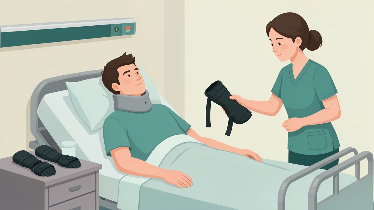
Stage 1 (0–6 weeks): resting hand splints at night hold the wrist in about 20–30° extension, MCP joints flexed 60–70°, IP joints extended and the thumb abducted.
Stage 2: Rehabilitation (6 weeks to about 6 months). Strengthen innervated muscles (deltoid, biceps, wrist extensors, serratus anterior, latissimus dorsi, pectoralis major) in graded, fatigue-aware programmes. Use 2–3 sets of 8–12 repetitions at about 60–70% of the one-repetition maximum, 3 times per week, adjusted for the shoulder-preservation guidelines (Consortium for Spinal Cord Medicine, 2005). Train transfers and wheelchair skills, and protect the shoulder from overuse.
Tenodesis preservation (all stages): Do not stretch the long finger flexors with the wrist in extension. Stretch the finger flexors into extension only with the wrist flexed, so that the flexors remain tight when the wrist extends. Passive finger flexion should be full with the wrist in flexion, and the thumb web space should be maintained.
Stage 3: Pre-surgical and post-surgical (from about 6–12 months). Surgery is usually considered after neurological plateau, commonly at least 6–12 months, once the patient has stable medical status, adequate passive ROM, motivation and realistic goals.
Tendon Transfer Principles and Donors
Transfer requires:
- Donor muscle of at least 4/5 (MRC), since a transferred muscle usually loses about one grade.
- Expendability: the donor loss must not cause functional loss.
- Full passive range in the recipient joint.
- Appropriate sensation and cognition.
- Synergy and a suitable direction of pull.
Elbow extension donors (important for C5–C6 patients):
- Posterior deltoid-to-triceps (Moberg): The posterior deltoid is attached to the triceps tendon via a graft (fascia lata or tibialis anterior). It requires a posterior deltoid of at least 4/5 and does not sacrifice elbow flexors. Postoperatively, the elbow is immobilised in full extension for about 4–6 weeks. Flexion is then gradually increased (about 20–30° per week) under protocol control, and the active extension programme begins with gravity-eliminated exercise. Avoid active flexion against resistance for 12 weeks, and avoid overstretching the repair.
- Biceps-to-triceps (Zancolli): The biceps is rerouted around the medial or lateral side of the humerus. It needs a biceps of at least 4/5 plus an intact brachialis and brachioradialis for elbow flexion. Postoperative splinting in extension lasts about 3 weeks, followed by a progressive flexion brace. This transfer is contraindicated by an elbow flexion contracture or deficient supination control, because the biceps is the primary forearm supinator. Elbow flexion contractures are a particular risk with this transfer.
Other donors: The brachioradialis is the standard donor for wrist extension (to ECRB) or key pinch in higher ICSHT groups. Other muscles with at least 4/5 strength are used to restore thumb flexion (flexor pollicis longus) and finger flexion.
Evidence and Clinical Pearls
- Elbow extension restored by surgery improves reach, transfer independence and pressure relief, and is associated with improved SCIM scores and quality of life (Hentz & Leclercq, 2002).
- The deltoid-to-triceps transfer may stretch out and lose effect if the splinting regimen is not followed.
- Tenodesis relies on tight finger flexors. A patient with C5 injury has no wrist extensors, so it cannot be used. Presence of tenodesis implies a C6 or greater wrist extensor function.
- A reported C7 "inability to extend against gravity" generally points to a C6 level or LMN involvement, so recheck the key muscle grades.
- Differentiate NLI, ZPP and functional level: the AIS A patient at C6 may still have an ICSHT group of 2–4 for planning reconstruction.
- Shoulder pain is common. Training the intact muscles should prioritise scapular stabilisers and avoid impingement positions.
Summary
Determine the NLI with ISNCSCI, using the lowest key muscle of at least 3/5 with the segment above normal. Map this to the functional level, which is the ICSHT group, and test for UMN versus LMN. Early care protects the spine, shoulders and hand posture, and preserves the tenodesis mechanism. Tendon transfer surgery, whose donors include the posterior deltoid and biceps, is considered once the neurology has stabilised. Donor strength should be at least 4/5 and passive range should be full.
References
- Rupp R, Biering-Sørensen F, Burns SP, et al. International Standards for Neurological Classification of Spinal Cord Injury: Revised 2019. Top Spinal Cord Inj Rehabil. 2021;27(2):1–22.
- Kirshblum SC, Burns SP, Biering-Sørensen F, et al. International standards for neurological classification of spinal cord injury (revised 2011). J Spinal Cord Med. 2011;34(6):535–546.
- Consortium for Spinal Cord Medicine. Preservation of upper limb function following spinal cord injury: a clinical practice guideline for health-care professionals. J Spinal Cord Med. 2005;28(5):434–470.
- McDowell CL, Moberg EA, House JH. The Second International Conference on Surgical Rehabilitation of the Upper Limb in Tetraplegia (Quadriplegia). J Hand Surg Am. 1986;11(4):604–608.
- Moberg E. Surgical treatment for absent single-hand grip and elbow extension in quadriplegia. J Bone Joint Surg Am. 1975;57(2):196–206.
- Bryden AM, Sinnott KA, Mulcahey MJ. Innovative strategies for improving upper extremity function in tetraplegia and considerations in measuring functional outcomes. Top Spinal Cord Inj Rehabil. 2005;10(4):75–93.
- Kalsi-Ryan S, Beaton D, Curt A, et al. The Graded Redefined Assessment of Strength, Sensibility and Prehension: reliability and validity. J Neurotrauma. 2012;29(5):905–914.
- Hentz VR, Leclercq C. Surgical Rehabilitation of the Upper Limb in Tetraplegia. London: WB Saunders; 2002.