This chapter introduces the structured process of clinical assessment and documentation, using acute mechanical low back pain and knee osteoarthritis as worked examples. It covers how findings are gathered, quantified, interpreted and recorded so that they support safe, defensible and reproducible care.
Why Assessment and Documentation Are Inseparable
Assessment generates data, clinical reasoning turns the data into hypotheses, and documentation preserves both so that other clinicians can reproduce and audit your decisions. Most physiotherapy systems use the ICF framework (body structure/function, activity, participation, plus personal and environmental factors) to organise findings. Documentation is a clinical, legal and communication tool. If something was not recorded, it is treated as not done.
Pathophysiology Relevant to the Index Cases
Acute mechanical low back pain (LBP). Lifting with a flexed, loaded spine can strain the lumbar paraspinal muscles or ligaments. It can also sensitise the facet joints or the outer annulus fibrosus of the disc. Pain arises from nociceptor activation, local inflammation and protective muscle guarding. Most episodes are non-specific, and about 90% improve substantially within 6 weeks. Phases are conventionally classed as acute (<6 weeks), subacute (6–12 weeks) and chronic (>12 weeks).
Knee osteoarthritis (OA). OA is a whole-joint disease. It involves progressive articular cartilage loss, subchondral bone remodelling, osteophyte formation, synovial inflammation and weakness of the periarticular muscles, especially the quadriceps. Synovitis produces effusion. Roughened articular surfaces produce crepitus. Radiographic severity is graded by Kellgren–Lawrence (KL) 0–4: 1 doubtful, 2 minimal osteophytes, 3 moderate narrowing, 4 severe narrowing with subchondral sclerosis. Radiographic grade correlates only weakly with pain and function.
Clinical Presentation
| Feature | Acute mechanical LBP (45-year-old female) | Knee OA (60-year-old male) |
|---|---|---|
| Onset | Sudden, after lifting | Insidious, progressive |
| Pain pattern | Mechanical: worse with flexion, eased by rest | Load-related; "gelling" |
| Morning stiffness | Variable | Typically <30 minutes |
| Neurological signs | None (no radicular symptoms) | None |
| Key findings | Limited flexion, paraspinal spasm | Crepitus, effusion, positive patellar grind, bony enlargement |
The ACR clinical criteria for knee OA include age >50, stiffness <30 minutes, crepitus, bony tenderness, bony enlargement and no palpable warmth. Palpable warmth, rapid onset of a hot swollen joint, fever or systemic illness should prompt consideration of septic or inflammatory arthritis, or crystal arthropathy.
Structured Subjective Assessment
Collect the following, and record it in the patient's own words where possible:
- Chief complaint, mechanism and onset
- Pain: location, quality, 24-hour behaviour, and severity on the Numeric Pain Rating Scale (NPRS, 0–10). The MCID is about 2 points.
- Aggravating and easing factors, and irritability (how easily symptoms provoke and how long they take to settle)
- Past medical history, medications, previous episodes, and goals
- Psychosocial "yellow flags": fear-avoidance, catastrophising, low mood, work dissatisfaction. The STarT Back Screening Tool stratifies LBP patients into low, medium or high risk of persistent disability.
Red Flag Screening (always documented, including when negative)
- Cauda equina syndrome: saddle anaesthesia, new urinary retention or incontinence, bilateral leg weakness. This needs emergency referral.
- Fracture: significant trauma, age >70, prolonged corticosteroid use, osteoporosis
- Malignancy: past cancer history, unexplained weight loss, night pain unrelieved by position
- Infection: fever, immunosuppression, recent infection or IV drug use
- Inflammatory disease: age <40 at onset, morning stiffness >30 minutes, improvement with exercise
- Vascular or visceral causes: pulsatile abdominal pain, pain unrelated to movement
Objective Examination: Measures and Normative Values
Observation and posture: record gait, swelling, muscle bulk, deformity (genu varum or valgum in knee OA) and antalgic posture.
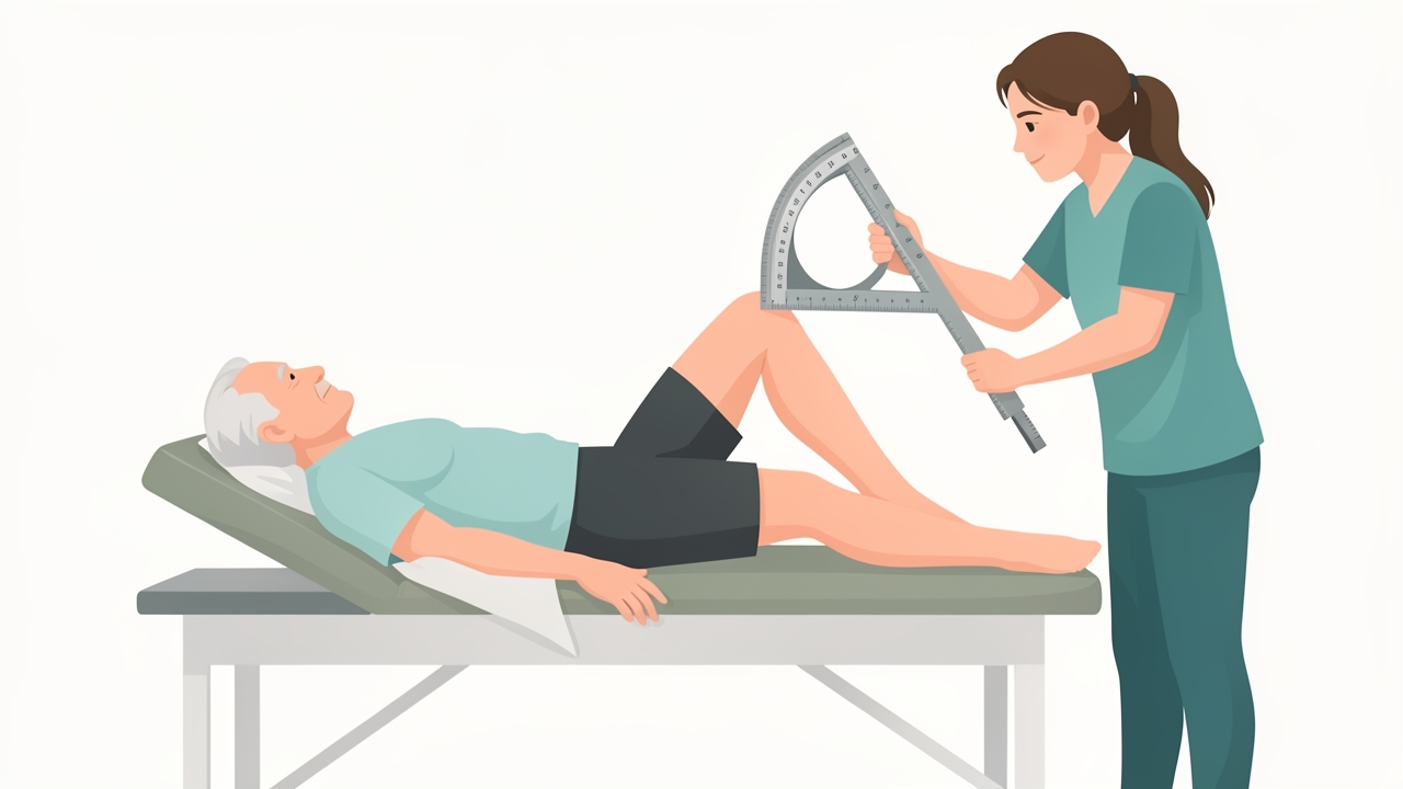
Record active and passive range with a standardised goniometer position. Normal knee flexion is 135–150°.
Range of motion (goniometry). Record active (AROM) and passive (PROM) values, end-feel and pain response. Use a standardised goniometer position and document it.
| Joint/Motion | Typical normative value |
|---|---|
| Lumbar flexion | ~60° (modified Schober: ≥5 cm increase on a 10 cm baseline) |
| Lumbar extension | ~25° |
| Lumbar lateral flexion | ~25° each side |
| Knee flexion | 135–150° |
| Knee extension | 0° (up to 5–10° hyperextension) |
Interpreting AROM versus PROM. In the knee OA case, crepitus with active flexion, effusion and a positive patellar grind point to articular involvement. PROM that is limited with a hard or capsular end-feel suggests joint pathology. In the knee, the capsular pattern is flexion limited more than extension. PROM that is greater than AROM, or that is full, suggests a muscular or neural contribution.
Strength. Use manual muscle testing on the MRC/Oxford 0–5 scale: 0 none, 1 flicker, 2 movement with gravity eliminated, 3 against gravity, 4 against moderate resistance, 5 normal. Where available, hand-held dynamometry gives objective quantification.
Neurological screen (lumbar). Test myotomes L2 hip flexion, L3 knee extension, L4 ankle dorsiflexion, L5 great toe extension and S1 plantarflexion. Test dermatomes and the patellar (L3–4) and Achilles (S1) reflexes. Perform the straight leg raise (SLR), which is positive when it reproduces radicular symptoms between about 30° and 70°, and the slump test. Document both sides.
Knee special tests.
- Effusion: stroke/bulge test (trace, 1+, 2+, 3+) and patellar tap
- Patellar grind (Clarke's) test: low specificity, so interpret alongside other findings
- Ligament and meniscal tests: Lachman, varus/valgus stress, McMurray. Include these when the history suggests instability or locking.
Functional performance and patient-reported outcome measures (PROMs):
- Oswestry Disability Index (ODI): 0–100%. Bands are 0–20 minimal, 21–40 moderate, 41–60 severe, 61–80 crippling and 81–100 bed-bound. The MCID is about 10 points.
- Roland–Morris Disability Questionnaire: 0–24, MCID about 5 points.
- WOMAC or KOOS for knee OA.
- OARSI-recommended performance tests: 30-second chair stand, 40 m fast-paced walk, and stair climb test.
- Timed Up and Go (TUG): ≥12 seconds suggests increased fall risk in older adults.
Reassess with the same measure, under the same conditions, at baseline and at intervals (for example every 2–4 weeks) so that change can be judged against measurement error and the MCID.
Clinical Reasoning and Diagnosis
Synthesise the data into a problem list and a primary hypothesis with differentials. For example: "Acute non-specific mechanical LBP with movement coordination impairment, no neurological deficit, low red flag risk, medium STarT Back risk." For the knee case: "Symptomatic tibiofemoral and patellofemoral OA (KL grade to be confirmed) with quadriceps weakness, effusion and activity limitation." Decide whether physiotherapy is appropriate or whether referral is needed. Record the reasoning that supports each decision.
Physiotherapy Management: Staged and Graded
Acute mechanical LBP
- Acute stage (0–2 weeks): reassurance and education that the prognosis is favourable. Advise early return to activity and avoiding bed rest. Offer pain-relieving positions and gentle, pain-limited mobility such as walking and pelvic tilts. Thermal modalities and manual therapy may give short-term relief. Teach lifting mechanics and pacing.
- Subacute stage (2–12 weeks): progress to motor control and trunk endurance work, such as bridging, bird-dog and side planks. Use directional-preference exercises if symptoms centralise. Add graded activity and work conditioning.
- Chronic or recurrent stage (>12 weeks): use a graded exercise programme, 2–3 sessions per week, with cognitive-behavioural principles and a return-to-work plan.
Contraindications and precautions: avoid spinal manipulation or end-range loading where there are red flags, suspected fracture, progressive neurological deficit or cauda equina signs.
Knee OA
- Education and self-management: load management, weight management (a loss of ≥5% body weight is beneficial in overweight patients), and footwear and walking aid advice.
- Strengthening: quadriceps and hip abductor/extensor exercises, 2–3 sessions per week, 2–3 sets of 8–12 repetitions at about 60–80% of 1RM. Progress from open-chain to closed-chain exercises (sit-to-stand, step-ups, mini-squats within a pain-tolerable range).
- Aerobic conditioning: aim for 150 minutes per week of moderate-intensity activity, such as walking, cycling or aquatic exercise. Include supervised exercise, land- or water-based tai chi, and balance training.
- Adjuncts: a cane in the contralateral hand, knee bracing for medial compartment loading, and TENS for short-term analgesia. Refer for medical review if effusion is persistent or an acute flare occurs.
Goal setting. Write goals in SMART form: Specific, Measurable, Achievable, Relevant, Time-bound. For example: "Patient will climb 12 stairs with NPRS ≤3/10 within 4 weeks," or "Improve ODI from 42% to ≤32% within 6 weeks."
Clinical Documentation Standards
Use a structured format such as SOAP (Subjective, Objective, Assessment, Plan), and make sure each entry includes:
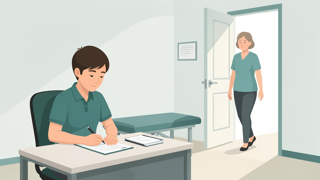
Use a structured format such as SOAP, with measurable, contemporaneous entries. If something was not recorded, it is treated as not done.
- Patient identifiers, date, time and the author's signature and designation
- Documented informed consent and any patient-reported adverse reactions
- Objective, measurable, unambiguous wording. Write "knee flexion AROM 95°, pain at end-range NPRS 6/10," not "limited ROM."
- Specific intervention dosage: exercise, sets, repetitions, load, frequency and duration
- Response to treatment and the plan for the next visit
- A clear record of education, home programme, referrals and discharge planning
- Corrections made by a single line through the error, with date and initials. Never use erasure.
- Contemporaneous entries, ideally completed the same day, in line with local record retention policy
Common errors include vague descriptors, copy-forward notes that do not reflect today's session, omission of negative findings, and missing goals or outcome measures.
Evidence and Clinical Pearls
- Current LBP guidelines favour education, staying active, and exercise and manual therapy over imaging, opioids or prolonged rest. Imaging is reserved for red flags or when results would change management.
- Radiographic OA severity does not predict symptoms or response to exercise. Treat the person, not the X-ray.
- Exercise therapy for knee OA is recommended by ACR and OARSI as a core treatment.
- Use the same PROM consistently, and interpret change against the MCID.
- Always test the joint above and below. Hip pathology can refer pain to the knee.
- Reassess the key finding after each intervention to judge whether it helped.
- Document negative findings: they show you considered and excluded serious pathology.
Summary
A competent assessment moves from a focused subjective history and red flag screen to an objective examination. This includes measured ROM, graded strength, neurological screening, special tests and validated outcome measures such as the NPRS, ODI, WOMAC and TUG. Findings are synthesised into a reasoned hypothesis and a staged, dosed treatment plan with SMART goals. Documentation must be accurate, measurable, timely and defensible. In acute LBP, the emphasis is on reassurance, early activity and graded exercise. In knee OA, it is on education, progressive strengthening, aerobic conditioning and weight management.
References
- George SZ, Fritz JM, Silfies SP, et al. Interventions for the management of acute and chronic low back pain: revision 2021. J Orthop Sports Phys Ther. 2021;51(11):CPG1–CPG60.
- Kolasinski SL, Neogi T, Hochberg MC, et al. 2019 American College of Rheumatology/Arthritis Foundation guideline for the management of osteoarthritis of the hand, hip, and knee. Arthritis Care Res. 2020;72(2):149–162.
- Bannuru RR, Osani MC, Vaysbrot EE, et al. OARSI guidelines for the non-surgical management of knee, hip, and polyarticular osteoarthritis. Osteoarthritis Cartilage. 2019;27(11):1578–1589.
- Altman R, Asch E, Bloch D, et al. Development of criteria for the classification and reporting of osteoarthritis: classification of osteoarthritis of the knee. Arthritis Rheum. 1986;29(8):1039–1049.
- Kellgren JH, Lawrence JS. Radiological assessment of osteo-arthrosis. Ann Rheum Dis. 1957;16(4):494–502.
- Fairbank JCT, Pynsent PB. The Oswestry Disability Index. Spine. 2000;25(22):2940–2953.
- Norkin CC, White DJ. Measurement of Joint Motion: A Guide to Goniometry. 5th ed. Philadelphia: F.A. Davis; 2016.
- Hill JC, Dunn KM, Lewis M, et al. A primary care back pain screening tool: identifying patient subgroups for initial treatment. Arthritis Rheum. 2008;59(5):632–641.