This chapter introduces the functional anatomy of the knee, the conditions most often seen in practice, and the structured, evidence-informed evaluation that links a patient's history and findings to an accurate diagnosis and a graded rehabilitation plan.
1. Why Systematic Knee Evaluation Matters
The knee is a high-load, low-congruency joint. It transmits about 2–3× body weight in walking and more than 7× in running, with little bony stability. Its stability depends on ligaments, menisci and muscle control, so injuries are common and often multi-structural.
Systematic evaluation serves four purposes:
- Screening for urgent pathology (fracture, septic arthritis, vascular injury).
- Differentiating mechanical, inflammatory and degenerative presentations.
- Establishing baselines with validated outcome measures.
- Guiding the choice and progression of treatment.
The standard sequence is history → observation → palpation → range of motion (ROM) → special tests. History generates hypotheses, and each later step confirms or refutes them. Special tests have modest accuracy in isolation and should only refine a diagnosis already suggested by the history.
2. Anatomy Overview
Articulations: The tibiofemoral joint is a modified hinge (bicondylar) joint. The patellofemoral joint is a sellar joint, and the proximal tibiofibular joint is a plane joint.
Menisci: The medial meniscus is C-shaped, less mobile and firmly attached to the deep MCL. The lateral meniscus is more circular and more mobile. Only the outer third (the red zone) is well vascularised, which has direct implications for healing and repair.
Ligamentous restraints:
| Structure | Primary restraint to |
|---|---|
| ACL (anteromedial and posterolateral bundles) | Anterior tibial translation; internal rotation |
| PCL | Posterior tibial translation |
| MCL (superficial and deep) | Valgus stress |
| LCL / posterolateral corner | Varus stress; external rotation |
| MPFL | Lateral patellar translation (0–30° flexion) |
Extensor mechanism: The quadriceps (including the vastus medialis obliquus, VMO), patellar tendon and patella. The iliotibial band (ITB) lies lateral to this mechanism.
Kinematics: The knee moves from 0° to about 135–140° of flexion, with 5–10° of passive hyperextension considered normal. The "screw-home" mechanism produces about 20° of external tibial rotation in terminal extension. Normal Q-angle is about 13–14° in males and 17–18° in females, and values above 20° are considered excessive.
3. Pathophysiology of Common Conditions
Ligament and meniscal injury. Non-contact pivoting, or a valgus force with the foot planted, loads the ACL, MCL and medial meniscus together. This is the classic "unhappy triad" described by O'Donoghue. Lateral meniscal tears actually accompany acute ACL rupture more often than medial ones. Medial tears become more common in chronic ACL deficiency.
Lateral patellar dislocation. This typically occurs with knee flexion, valgus and femoral internal rotation during a pivot. The MPFL tears, usually at its femoral attachment. Predisposing factors include trochlear dysplasia, patella alta, a TT–TG distance above 20 mm, ligamentous laxity and female sex.
Iliotibial band syndrome (ITBS). Compression of richly innervated fat and connective tissue deep to the ITB occurs near the lateral femoral epicondyle (Fairclough et al., 2006). It is most provocative around 20–30° of flexion, which corresponds to foot strike and downhill running.
Patellofemoral pain (PFP). This is multifactorial, with overload of the patellofemoral joint from hip, knee and foot mechanics and from training load errors.
Osteoarthritis (OA). Degenerative cartilage loss with subchondral remodelling and osteophytes. It is mechanically driven and often medial-compartment dominant.
Rheumatoid arthritis (RA). An autoimmune synovitis that produces symmetrical, inflammatory joint involvement.
4. Clinical Presentation: Pattern Recognition
| Patient | Key features | Leading hypothesis |
|---|---|---|
| 19 F basketball, pivot | Visible displacement or spontaneous reduction, medial pain, apprehension | Lateral patellar dislocation (MPFL injury) |
| 22 M soccer, valgus force | Medial pain, instability, effusion | ACL + medial meniscus + MCL (unhappy triad) |
| 28 M footballer, "pop" | Swelling within about 2 hours | Haemarthrosis, most commonly ACL rupture (about 70% of acute traumatic haemarthroses) |
| 32 runner, lateral pain | Pain at about 30° flexion, downhill running | ITBS |
| 40 M, stairs and "movie sign" | Anterior pain, worse with sitting | PFP |
| 45 F, bilateral pain | Morning stiffness of 90 min, raised ESR | Inflammatory arthritis (RA) |
| 55 M, medial pain | Joint space narrowing, stiffness of 20 min | Medial tibiofemoral OA |
Swelling timing is diagnostic. Swelling within 2 hours implies intra-articular bleeding (ACL, peripheral meniscal tear, osteochondral fracture, patellar dislocation). Swelling that develops over 12–24 hours suggests synovial effusion, as in a meniscal tear or irritated OA.
Morning stiffness discriminates between inflammatory and degenerative disease. Stiffness lasting more than 60 minutes suggests inflammatory disease. Stiffness under 30 minutes suggests OA, which fits the ACR clinical criteria for knee OA.
5. Red Flags and Screening
Refer or investigate urgently for:
- Ottawa Knee Rule positives: age ≥55, isolated patellar tenderness, fibular head tenderness, inability to flex to 90°, or inability to bear weight for four steps (Stiell et al., 1996).
- Hot, swollen joint with fever: exclude septic arthritis.
- Gross deformity or multiligament instability: suspect knee dislocation and check pedal pulses and ankle–brachial index (popliteal artery injury).
- True locked knee: a mechanical block to extension, suggesting a displaced bucket-handle tear.
- Calf pain, swelling or warmth: apply the Wells score for DVT.
- Night or constant pain, unexplained weight loss or past malignancy: consider tumour.
- Foot drop or numbness: consider common peroneal nerve injury.
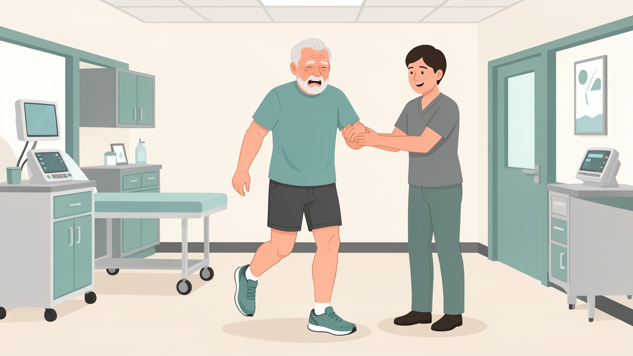
Ottawa Knee Rule positives, such as age 55 or over or being unable to bear weight for four steps, need urgent referral or investigation.
6. Assessment: Measures and Normative Values
Observation. Look at gait, alignment (genu varum or valgum), effusion, quadriceps atrophy and patellar position.
Palpation. Check joint line tenderness, patellar facets, the MCL and Gerdy's tubercle for ITB, and use temperature comparison.
Effusion grading. The stroke test grades effusion from zero to trace, 1+, 2+ or 3+. The patellar tap suggests large effusions.
ROM. Measure with a goniometer. Normal values are 0° extension (to 5–10° hyperextension) and 135–140° flexion. Functional needs are about 65° for gait, 90° for stairs, 105° for rising from a chair and over 110° for kneeling.
Strength. Use manual muscle testing (MRC/Oxford 0–5) or handheld dynamometry. Isokinetic or dynamometer limb symmetry index (LSI) ≥90% is a common return-to-sport threshold.
Special tests:
| Test | Position and finding |
|---|---|
| Lachman | 20–30° flexion; anterior translation with a soft endpoint. The most sensitive ACL test (about 85%). |
| Pivot shift | Very specific for ACL (about 98%), but difficult in an awake patient |
| Valgus stress | At 0° (posteromedial capsule and cruciates) and 30° (isolated MCL); laxity grade I 0–5 mm, II 5–10 mm, III >10 mm |
| Posterior sag/drawer | PCL |
| McMurray / Thessaly (20°) | Meniscal tear; moderate accuracy, best interpreted with joint line tenderness |
| Patellar apprehension | MPFL insufficiency |
| Noble compression | Pain at about 30° flexion with ITB pressure; ITBS |
Patient-reported outcome measures (PROMs): KOOS, IKDC-subjective, Lysholm, Tegner (activity), Kujala/AKPS (PFP), WOMAC (OA) and LEFS.
Performance measures: 30-second chair stand, Timed Up and Go, 6-minute walk test, single-leg hop battery (LSI ≥90%) and step-down quality.
Imaging and labs: Radiographs are graded by Kellgren–Lawrence (0–4). Use MRI for suspected soft-tissue injury when the result would change management. ESR, CRP, rheumatoid factor and anti-CCP support suspected RA.
7. Physiotherapy Management: Staged Principles
Stage 1: Protection and symptom control (days 0–14)
- Use POLICE (protection, optimal loading, ice, compression, elevation).
- Use crutches and a hinged brace as needed, with weight-bearing as tolerated if no fracture is present.
- Perform quadriceps setting, straight-leg raises, heel slides and ankle pumps.
- The ROM goal is full extension and about 90° flexion.
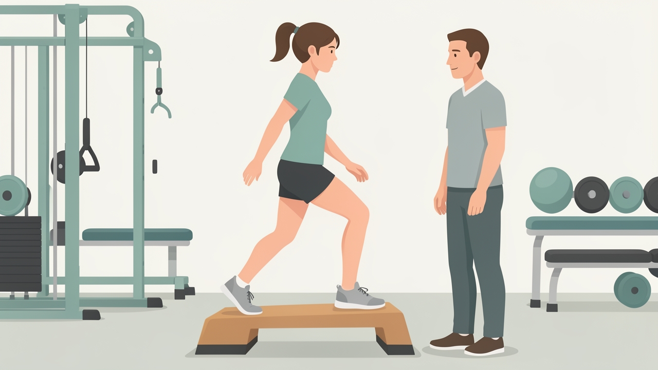
Stage 2 (weeks 2–6): progress to closed-chain work such as mini-squats, step-ups and bike.
Stage 2: Restoring ROM and neuromuscular control (weeks 2–6)
- Progress to closed-chain work: mini-squats, step-ups and bike.
- Begin balance and proprioception training.
- Add hip abductor and extensor strengthening.
Stage 3: Strengthening (weeks 6–12+)
- Dose at 2–3 sessions per week, 2–4 sets of 8–12 repetitions at 60–80% 1RM, progressing to heavier loads (about 6 reps) for hypertrophy and strength.
- Include eccentric control and posterior chain work.
Stage 4: Return to sport (about 6–12 months for ACL)
- Progress through running, plyometrics, cutting and sport-specific drills.
- Criteria-based discharge: LSI ≥90% on strength and hop tests, good movement quality, psychological readiness (ACL-RSI) and no effusion. Delaying return to sport until about 9 months reduces re-injury (Grindem et al., 2016).
8. Condition-Specific Management
First-time patellar dislocation. Manage non-operatively with early protected motion, a patellar-stabilising brace for 2–4 weeks if needed, and quadriceps and hip strengthening. Surgery (MPFL reconstruction) is considered for recurrent instability or osteochondral fracture. Avoid early aggressive end-range flexion with lateral loading.
ACL / multiligament injury. Choose between structured non-operative rehabilitation and reconstruction according to age, activity goals and instability. Prehabilitation (restore ROM, reduce effusion, build strength) improves outcomes. Isolated grade I–II MCL sprains heal well non-operatively.
ITBS. Modify running volume (especially downhill), strengthen hip abductors (side-lying abduction, side plank, single-leg squat), and consider cadence and gait retraining. Stretching alone has limited evidence.
PFP. Use combined hip and knee exercise therapy as first-line care, with taping or foot orthoses as short-term adjuncts and load management (Crossley et al., 2016).
OA. Education, exercise (about 12 weeks, 2–3 sessions/week, plus 150 min/week of aerobic activity) and weight loss if overweight are core treatments, consistent with the ACR/AF 2019 guideline (Kolasinski et al., 2020). Strongly recommended options include tai chi, walking and aquatic exercise. Walking aids and knee bracing can be considered according to compartment involvement.
RA. Refer early to rheumatology for DMARD therapy within about 3 months of symptom onset. Physiotherapy addresses joint protection, graded strengthening and aerobic work, with exercise adjusted during flares.
9. Evidence and Clinical Pearls
- No single special test rules diagnoses in or out. Cluster findings (history + joint line tenderness + provocation tests).
- A rapidly swelling knee after a "pop" is an ACL tear until proven otherwise.
- Always compare with the uninjured side, and test the Lachman and valgus stress with the patient relaxed.
- Examine the hip and ankle: abnormal hip control commonly drives knee symptoms.
- Pain at about 30° flexion on the lateral side points to ITBS, not meniscal pathology.
- The "movie sign" (pain on prolonged sitting with flexed knees) is typical of PFP.
- Bilateral symptoms with prolonged morning stiffness warrant laboratory investigation, not just exercise.
- Radiographic OA severity correlates poorly with pain and function, so treat the person, not the X-ray.
- Reassess regularly with the same PROM and performance measures to demonstrate change. MCID varies by instrument, for example roughly 8–10 points for KOOS subscales.

The "movie sign", pain on prolonged sitting with flexed knees, is typical of patellofemoral pain.
10. Summary
Effective knee evaluation combines anatomical knowledge, pattern recognition from the history, systematic examination, and validated measures. Mechanism and swelling timing guide hypotheses about the injured structure. Morning stiffness separates inflammatory from degenerative disease. Special tests confirm rather than create diagnoses, and red flags must be excluded first. Physiotherapy management follows staged, criteria-based progression, with exercise as the cornerstone for most conditions, and outcomes are tracked with PROMs and objective performance measures.
References
- Magee DJ. Orthopedic Physical Assessment. 7th ed. St. Louis: Elsevier; 2020.
- Cook C, Hegedus E. Orthopedic Physical Examination Tests: An Evidence-Based Approach. 2nd ed. Upper Saddle River, NJ: Pearson; 2013.
- Stiell IG, Greenberg GH, Wells GA, et al. Prospective validation of a decision rule for the use of radiography in acute knee injuries. JAMA. 1996;275(8):611-615.
- Kolasinski SL, Neogi T, Hochberg MC, et al. 2019 American College of Rheumatology/Arthritis Foundation guideline for the management of osteoarthritis of the hand, hip, and knee. Arthritis Care Res. 2020;72(2):149-162.
- Crossley KM, van Middelkoop M, Callaghan MJ, et al. 2016 Patellofemoral pain consensus statement from the 4th International Patellofemoral Pain Research Retreat, Manchester. Part 2: recommended physiotherapy management. Br J Sports Med. 2016;50(14):844-852.
- Grindem H, Snyder-Mackler L, Moksnes H, Engebretsen L, Risberg MA. Simple decision rules can reduce reinjury risk by 84% after ACL reconstruction: the Delaware-Oslo ACL cohort study. Br J Sports Med. 2016;50(13):804-808.
- Aletaha D, Neogi T, Silman AJ, et al. 2010 Rheumatoid arthritis classification criteria: an ACR/EULAR collaborative initiative. Arthritis Rheum. 2010;62(9):2569-2581.
- Fairclough J, Hayashi K, Toumi H, et al. The functional anatomy of the iliotibial band during flexion and extension of the knee: implications for understanding iliotibial band syndrome. J Anat. 2006;208(3):309-316.