1.1 The Kinetic Chain Concept: A Foundational Principle
The kinetic chain (KC) describes the coordinated, interdependent sequencing of movement across multiple joints and segments, where motion at one joint influences motion at adjacent and distal joints. Originally described by Steindler (1955) in the context of biomechanics, the concept has become central to understanding human movement, particularly in rehabilitation and sports performance. The KC is often conceptualized as a series of rigid segments (bones) connected by joints, with muscles acting as active stabilizers and movers. Energy is transferred proximally to distally, allowing efficient force production and dissipation. In the upper extremity, the kinetic chain originates from the lower limbs and core, passing through the trunk, scapula, and glenohumeral joint to the hand. Disruption at any point—whether due to weakness, inflexibility, or motor control deficits—can lead to compensatory patterns and increased injury risk, especially at the shoulder.
1.2 Open vs. Closed Kinetic Chain: Clinical Distinctions
Kinetic chain exercises are classified as open or closed based on the relationship of the distal segment to a fixed surface.
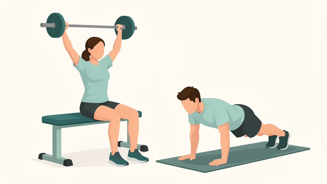
Open chain: the hand moves freely, as in a seated dumbbell press. Closed chain: the hand is fixed, as in a push-up, loading the shoulder with more compression and less shear.
- Open Kinetic Chain (OKC): The distal segment (e.g., hand or foot) moves freely in space. Examples include bicep curls, shoulder flexion in the sagittal plane, and seated dumbbell press. OKC exercises typically isolate a single joint and muscle group, making them useful for targeted strengthening but less functional for multiplanar activities. They often produce greater shear forces at the glenohumeral joint, which can be problematic in unstable shoulders.
- Closed Kinetic Chain (CKC): The distal segment is fixed (e.g., hand on a wall or floor), and movement occurs at proximal joints. Examples include push-ups, wall slides, and planks. CKC exercises promote co-contraction of agonist and antagonist muscles, enhancing joint stability and proprioception. They load the joint in a more compressive manner, reducing shear and often safer for rehabilitation. However, CKC exercises may not adequately challenge the rotator cuff in isolation.
Clinical Relevance: The choice between OKC and CKC depends on the phase of rehabilitation, patient goals, and pathology. For example, early post-operative rotator cuff repair often emphasizes CKC to minimize shear, while late-phase return to throwing incorporates OKC for power development.
1.3 Shoulder Biomechanics: An Integrated System
The shoulder complex comprises four joints: glenohumeral (GH), acromioclavicular (AC), sternoclavicular (SC), and scapulothoracic (ST). The GH joint is a ball-and-socket with high mobility but low inherent stability, relying on dynamic stabilizers (rotator cuff, scapular muscles) and static stabilizers (labrum, capsule, ligaments). The scapula serves as a critical link between the trunk and the humerus, providing a mobile base for glenohumeral motion. Normal shoulder function requires coordinated motion between these joints, known as scapulohumeral rhythm.
1.4 Scapulohumeral Rhythm: The 2:1 Ratio
Scapulohumeral rhythm (SHR) describes the coordinated movement between the scapula and humerus during arm elevation. In healthy individuals, for every 2° of GH abduction, the scapula upwardly rotates approximately 1°, giving a 2:1 ratio (GH:ST). This ratio is not fixed and varies with plane of motion, load, and individual anatomy. During the first 30° of abduction, scapular motion is variable (setting phase), followed by a consistent 2:1 ratio up to 120°, after which the ratio may decrease. Disruption of SHR—such as scapular dyskinesis—can lead to subacromial impingement, rotator cuff pathology, and glenohumeral instability.
Measurement Protocol: Scapular upward rotation can be measured using a digital inclinometer or motion capture. Clinically, the scapular assistance test (SAT) and scapular retraction test (SRT) help assess the impact of scapular position on shoulder symptoms.
1.5 Glenohumeral Stability Mechanisms
Glenohumeral stability is maintained by a combination of passive and active mechanisms:
- Passive: Bony geometry (glenoid version, humeral head curvature), labrum (deepens socket), capsuloligamentous structures (inferior glenohumeral ligament, coracohumeral ligament), and negative intra-articular pressure.
- Active: Rotator cuff muscles (supraspinatus, infraspinatus, teres minor, subscapularis) compress the humeral head into the glenoid (concavity-compression). The deltoid and biceps brachii also contribute. Scapular muscles (trapezius, serratus anterior) position the glenoid optimally for humeral articulation.
Assessment Parameters: The apprehension test, relocation test, and sulcus sign are used to assess instability. In a clinical setting, measuring posterior capsular tightness (e.g., cross-body adduction test) and rotator cuff strength (e.g., using a handheld dynamometer) provides objective data.
1.6 The Kinetic Chain in Overhead Throwing: A Model of Energy Transfer
The overhead throwing motion exemplifies kinetic chain function. The lower extremities generate force during the stride and hip rotation, which is transferred through the trunk and pelvis (core) to the shoulder and arm. The sequence is: hip and trunk rotation → scapular retraction and upward rotation → glenohumeral external rotation and acceleration → wrist and finger flexion. Any delay or weakness in the proximal segments forces the shoulder to compensate, increasing stress on the rotator cuff and labrum. This is known as "catch-up" phenomenon.

Force for the arm starts in the legs and hips and passes through the trunk to the shoulder; any delay or weakness lower in the chain makes the shoulder compensate.
Clinical Measurements: To assess kinetic chain efficiency, clinicians use the Y-Balance Test (Upper Quarter) and the Closed Kinetic Chain Upper Extremity Stability Test (CKCUEST). The CKCUEST involves performing as many hand touches as possible in 15 seconds, with a distance of 36 inches (91.4 cm) between hands. Normative values vary by sex and sport; a score below 21 touches is considered poor in male athletes (Tucci et al., 2020).
1.7 Assessment Parameters for Kinetic Chain Dysfunction
A comprehensive assessment of the kinetic chain in shoulder patients includes:
- Posture: Forward head, rounded shoulders, thoracic kyphosis (upper crossed syndrome).
- Scapular Dyskinesis: Visual observation during arm elevation (scapular winging, inferior angle prominence).
- Core Stability: Plank test, side plank endurance, and McGill’s core stability tests.
- Hip and Trunk Range of Motion: Hip internal/external rotation, trunk rotation (using goniometer).
- Muscle Strength: Lower trapezius, serratus anterior, and rotator cuff via manual muscle testing or dynamometry.
- Neuromuscular Control: Single-leg stance, star excursion balance test.
Protocol Example: The Scapular Dyskinesis Test (SDT) involves 5 repetitions of bilateral arm elevation in the scapular plane. A positive test is the presence of dysrhythmia, winging, or asymmetrical movement.
1.8 Treatment Approaches: Restoring Kinetic Chain Function
Rehabilitation should address all links in the chain, progressing from proximal to distal stability and then to functional integration.
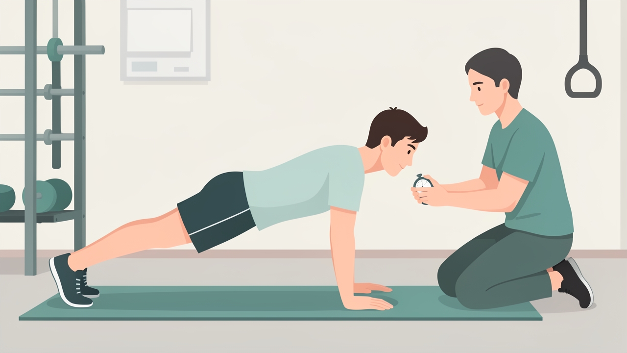
Proximal stabilization comes first: core and hip work such as planks, building to 60-second holds for endurance.
- Proximal Stabilization: Core and hip strengthening (e.g., dead bugs, planks, clamshells, hip bridges). Focus on endurance—hold times of 60 seconds for planks.
- Scapular Control: Scapular retraction exercises (rows, prone Ys and Ts), serratus anterior activation (push-up plus, wall slides). Use of biofeedback (e.g., mirror, EMG) to correct dyskinesis.
- Glenohumeral Strengthening: Rotator cuff exercises in neutral and elevated positions. Begin with isometrics, then progress to isotonic (theraband, dumbbells). Avoid painful arcs.
- Neuromuscular Re-education: Closed kinetic chain exercises (e.g., rhythmic stabilization, ball on wall). Progress to open chain with perturbations.
- Sport-Specific Training: Integrate kinetic chain drills (e.g., medicine ball throws, cable rotations) with emphasis on timing and sequencing.
Example Protocol for Throwing Shoulder (Phase 1):
- Week 1-2: Scapular setting, sidelying external rotation (0° abduction), supine serratus punches, core stability (dead bug).
- Week 3-4: Prone Y/T/W, standing rows, push-up plus, and lower body strengthening (lunges, squats).
- Week 5-6: Begin CKCUEST, light medicine ball throws, and progress to eccentric rotator cuff.
1.9 Clinical Pearls and Evidence-Based Practice
- Measurement of Scapular Upward Rotation: Use a digital inclinometer at the scapular spine with the arm at 0°, 90°, and 120° of abduction. Normal values: ~10° at rest, ~40° at 120° abduction.
- CKCUEST Normative Data: For healthy young adults, average touches: males 25±5, females 21±4 (Tucci et al., 2020). A side-to-side asymmetry >10% may indicate injury risk.
- Kinetic Chain Deficits in Shoulder Pain: Patients with subacromial impingement often exhibit decreased hip extension and trunk rotation ROM compared to controls (Kibler et al., 2013).
- Treatment Progression: Always address proximal deficits first. A weak core cannot generate sufficient force for overhead activities, forcing the shoulder to overload.
1.10 Conclusion
Understanding the kinetic chain and shoulder biomechanics is essential for effective diagnosis and treatment of shoulder pathologies. The shoulder does not function in isolation; it is a link in a complex chain that begins in the lower extremities. Clinicians must assess and treat the entire kinetic chain, not just the symptomatic joint. By integrating measurements like scapular upward rotation, CKCUEST, and core endurance tests, practitioners can identify underlying dysfunctions and design targeted, evidence-based rehabilitation programs. The interplay of open and closed kinetic chain exercises, combined with a focus on neuromuscular control, allows for a comprehensive approach that reduces injury risk and optimizes performance.
References
- Steindler, A. (1955). Kinesiology of the human body under normal and pathological conditions. Charles C Thomas.
- Kibler, W. B., Ludewig, P. M., McClure, P. W., Michener, L. A., Bak, K., & Sciascia, A. D. (2013). Clinical implications of scapular dyskinesis in shoulder injury: the 2013 consensus statement from the 'Scapular Summit'. British Journal of Sports Medicine, 47(14), 877–885. https://doi.org/10.1136/bjsports-2013-092425
- Tucci, H. T., Martins, J., Sposito, G., & Camarini, P. M. F. (2020). Closed kinetic chain upper extremity stability test (CKCUEST) in healthy young adults: normative values and reliability. Brazilian Journal of Physical Therapy, 24(6), 512–519. https://doi.org/10.1016/j.bjpt.2019.09.002
- McQuade, K. J., & Smidt, G. L. (1998). Dynamic scapulohumeral rhythm: the effects of external resistance during elevation of the arm in the scapular plane. Journal of Orthopaedic & Sports Physical Therapy, 27(2), 125–133. https://doi.org/10.2519/jospt.1998.27.2.125
- Ellenbecker, T. S., & Cools, A. (2010). Rehabilitation of shoulder impingement syndrome and rotator cuff injuries: an evidence-based review. British Journal of Sports Medicine, 44(5), 319–327. https://doi.org/10.1136/bjsm.2009.059923
- Wilk, K. E., Arrigo, C. A., & Andrews, J. R. (1997). Current concepts: the stabilizing structures of the glenohumeral joint. Journal of Orthopaedic & Sports Physical Therapy, 25(6), 364–379. https://doi.org/10.2519/jospt.1997.25.6.364
- Sciascia, A., & Cromwell, R. (2012). Kinetic chain rehabilitation: a theoretical framework. International Journal of Sports Physical Therapy, 7(4), 400–413. https://www.ncbi.nlm.nih.gov/pmc/articles/PMC3414076/