This chapter introduces blood flow restriction (BFR) therapy as a rehabilitation tool: its definition and history, its physiological mechanisms, how to screen and assess patients, how to set pressure and exercise parameters, and how to progress safely.
Definition and History of BFR Therapy
BFR therapy applies a pneumatic cuff or tourniquet to the proximal portion of a limb during exercise. The cuff partially restricts arterial inflow and substantially restricts venous outflow from the working muscle. Venous return is reduced rather than reliably abolished, and cuff width, pressure and limb size all influence this. The result is blood pooling distal to the cuff and a metabolically demanding environment, even at very low external loads.
The technique originated with Dr. Yoshiaki Sato in Japan, who conceived "KAATSU" training in 1966 and refined it over the following decades. Early research (Takarada, Ishii and colleagues, around 2000) showed that low-load exercise with vascular restriction produced strength and size gains. The method moved into Western rehabilitation after 2010, and a consensus on safety and application was published in 2019 (Patterson et al.).
Physiological Mechanisms of BFR
Heavy loading (≥70% 1RM) drives hypertrophy through mechanical tension. BFR achieves comparable adaptation at 20–30% 1RM through several overlapping mechanisms:
- Metabolic stress. Trapped, hypoxic muscle accumulates lactate, H⁺ and inorganic phosphate. These stimulate group III/IV afferents, which raises sympathetic drive and perceived effort.
- Early fast-twitch recruitment. Hypoxia and fatigue of slow-twitch units force recruitment of higher-threshold motor units despite a light load. EMG amplitude is higher than with unrestricted exercise at the same load.
- Cellular swelling. Fluid shifts into the muscle cell and acts as an anabolic signal.
- Anabolic signalling. mTORC1 and S6K1 phosphorylation increase muscle protein synthesis. Myostatin expression falls and satellite-cell activity rises.
- Systemic hormonal response. Growth hormone rises acutely, but its contribution to local hypertrophy is probably small.
- Exercise-induced hypoalgesia. Pain often falls after BFR exercise, which is useful in painful knees.
Early strength gains (first 2–4 weeks) are partly neural. Measurable hypertrophy generally follows after 3–6 weeks.
Clinical Presentation of Suitable Candidates
BFR suits patients who cannot tolerate or safely perform high-load training, or who have plateaued with conventional loading. Typical presentations include:
- Post-operative atrophy. Examples are ACL reconstruction at 2–4 weeks, partial meniscectomy at 8 weeks, and arthroscopic debridement. These patients show quadriceps atrophy, extensor lag and arthrogenic muscle inhibition.
- Patellofemoral pain syndrome. Anterior knee pain on squatting or stairs, VMO weakness and a positive patellar grind test. High-load squatting provokes pain, so the aim is quadriceps strengthening with low joint stress.
- Tendinopathy. Examples are patellar tendinopathy and Achilles tendinopathy, where heavy loading may be irritable. BFR can supplement or substitute for loading during the reactive phase.
- Strength plateau. An athlete 6 months after ACLR whose quadriceps limb symmetry index (LSI) has stalled despite conventional low-load work.
Screening, Contraindications and Red Flags
Screening must precede any cuff application.
Absolute contraindications:
- Previous deep vein thrombosis or pulmonary embolism, or active VTE
- Severe peripheral vascular disease, arterial grafts or revascularisation in the limb
- Limb lymphedema or dialysis access in the limb
- Uncontrolled hypertension (resting BP above approximately 180/110 mmHg)
- Sickle cell disease or trait with crisis history
- Active infection, open unhealed wounds under the cuff, or suspected compartment syndrome
- Pregnancy (limb application is generally avoided)
- Active malignancy and rhabdomyolysis
Relative contraindications:
- Controlled hypertension
- Diabetes with neuropathy or microvascular disease
- Obesity
- Smoking
- Recent surgery with fresh incisions
- Clotting-risk medication or oral contraceptive use
- Family history of VTE
- Fragile skin or severe anxiety
Screening tools: resting BP and heart rate, a Wells DVT score if there is any suspicion of calf pain or swelling, a Caprini or equivalent VTE risk assessment post-operatively, and a peripheral pulse and capillary refill check.
Red flags during or after exercise: unilateral calf pain, swelling, warmth or redness; chest pain or dyspnoea; syncope or pre-syncope; persistent numbness or pallor; severe pain disproportionate to effort; dark urine (rhabdomyolysis); and unexplained petechiae or bruising.
Reported adverse-event rates are low. A large Japanese survey found DVT in roughly 0.05% and rhabdomyolysis in around 0.01% of users (Nakajima et al., 2006). Rates are lower when pressures are individualised and exercise is supervised.
Assessment: Baseline and Outcome Measures
Record baseline measures before starting, and repeat them every 2–4 weeks.
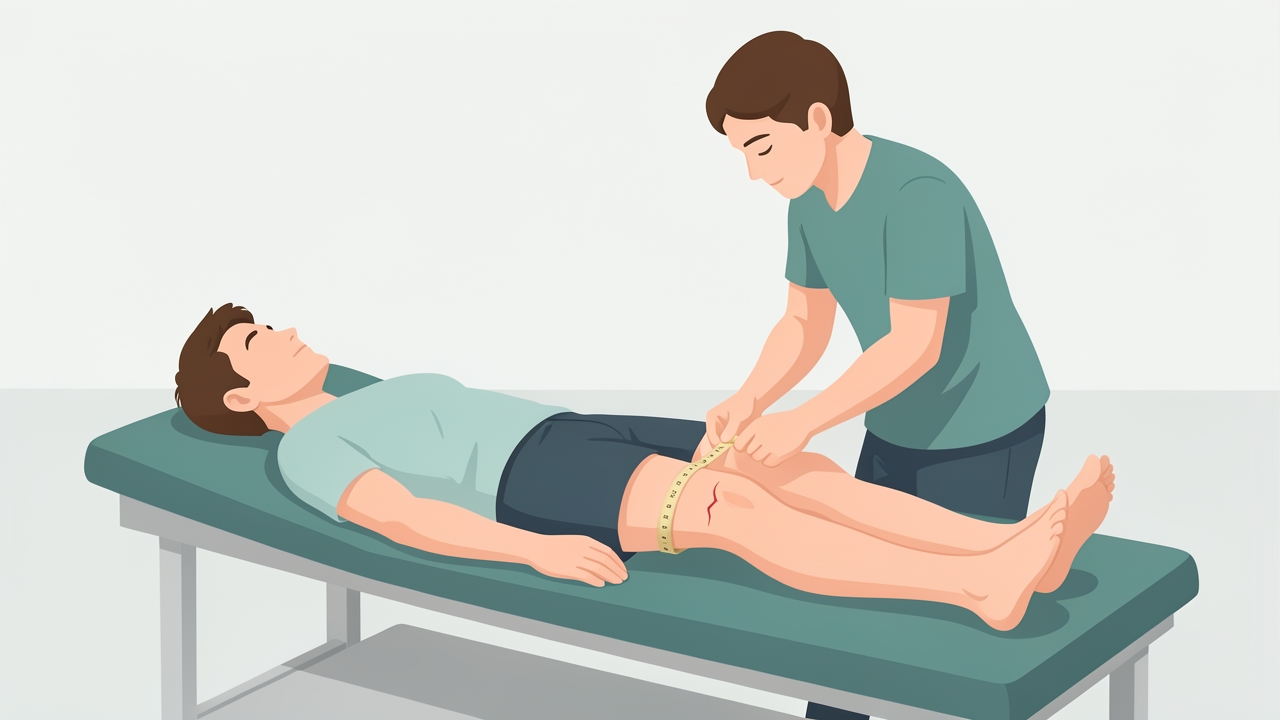
Thigh girth is taken 10 cm and 15 cm above the superior pole of the patella. A difference above 1–2 cm indicates clinically meaningful atrophy.
- Range of motion (goniometry). Normal knee extension is 0° (0–5° hyperextension) and flexion is 135–140°. Measure any extensor lag, in degrees, during a supine active straight leg raise.
- Strength. Use manual muscle testing (MMT, Oxford 0–5). Better still, use handheld dynamometry or isokinetic testing at 60°/s. Compute LSI as involved ÷ uninvolved × 100. Return-to-sport thresholds are typically ≥90%.
- Girth. Take thigh circumference 10 cm and 15 cm above the superior pole of the patella. Girth differences above 1–2 cm indicate clinically meaningful atrophy.
- Function. Use the 30-second chair stand, step-down test, single-leg squat quality and the single-leg hop battery (single, triple, crossover, 6 m timed). Hop LSI should be ≥90%.
- Patient-reported outcomes. Use KOOS or IKDC for post-surgical patients and the Kujala score for patellofemoral pain (100 = normal; below about 85 suggests clear dysfunction). Use VISA-P for patellar tendinopathy (100 = asymptomatic) and LEFS for general lower-limb function. Record pain on an NPRS from 0 to 10.
- Special tests. The patellar grind (Clarke's) test and palpation help identify patellofemoral involvement.
Pressure Settings and BFR Parameters
Limb occlusion pressure (LOP) is the minimum pressure that stops arterial inflow at the cuff site. Measure it individually with a Doppler probe or an integrated cuff sensor. Do not use a fixed arbitrary pressure. Typical thigh LOP is about 150–250 mmHg with a 10–12 cm cuff, but it varies with limb circumference, BP, cuff width and cuff type.
Prescribe pressure as a percentage of LOP:
| Application | Typical % LOP |
|---|---|
| Lower limb exercise | 60–80% |
| Upper limb exercise | 40–60% |
| Elastic (wrap) cuffs, subjective | ~7/10 tightness |
Example: if LOP is 200 mmHg and the target is 80%, set the cuff to 160 mmHg.
Pressure also depends on cuff width. Wide cuffs (10–12 cm) occlude at lower pressures than narrow cuffs (5 cm), so the cuff must always be matched to the limb and the pressure calculated from that cuff. Position the cuff at the proximal thigh (inguinal fold). Cuff pressure is typically set to LOP measured with the same cuff, with the patient in the working position.
Exercise prescription (standard protocol):
- Load: 20–30% of 1RM (or a pain-free equivalent when 1RM testing is unsafe)
- Repetitions: 30-15-15-15 (75 total) per exercise
- Rest: 30–60 seconds between sets, with the cuff inflated
- Tempo: about 1–2 seconds concentric and eccentric
- Frequency: 2–3 sessions per week; in early post-operative rehabilitation up to daily short bouts are used
- Duration: 4–8 weeks typically
- Cuff time: keep continuous inflation under about 20 minutes, deflating between exercises as needed
- Effort: sets are completed close to, but not necessarily to, failure
A patient who asks "how light is light?" can be told that for a leg extension with a 1RM of 60 kg, 30% is roughly 18 kg. For the athlete in the first stage of rehab, an even lighter load, or bodyweight, is acceptable.
Physiotherapy Management: A Staged Approach
Stage 1: Early post-operative or irritable phase (weeks 0–3). Aim to limit atrophy and control pain. Apply BFR with the cuff at 50–80% LOP using low-intensity activities such as quadriceps sets, straight leg raises and heel slides, if surgeon clearance and wound status allow. Perform 4 sets (30-15-15-15) with 30 seconds' rest, once or twice daily. Add neuromuscular electrical stimulation if inhibition is marked.
Stage 2: Low-load strengthening (weeks 3–8). Progress to open and closed chain exercises at 20–30% 1RM: leg press, leg extension (within the surgeon's protocol for ACLR), mini squats and step-ups. Use 60–80% LOP, 30-15-15-15, 2–3 times per week. Increase load only when exercises are pain-free (NPRS ≤3/10).
Stage 3: Blended loading (weeks 8–16). Combine BFR sessions with progressive heavier non-BFR training (60–80% 1RM) on separate days. Reserve BFR for athletes who plateau, who have joint irritability or who need extra training volume.
Stage 4: Return to sport. Taper BFR as LSI reaches 90% on strength and hop tests. Maintain heavy strength work and add plyometrics and sport-specific loading.
Special cases:
- Patellar tendinopathy: use BFR with isometric or low-load knee extension during reactive phases, then transition to heavy slow resistance training.
- Patellofemoral pain: use BFR leg extension in a pain-free range (typically 90–45° flexion) and closed-chain work at limited depth.
- Post-meniscectomy: follow weight-bearing and ROM restrictions; BFR can begin once the wound is healed.
Monitoring: record perceived exertion (RPE) and discomfort every set, and check skin colour and distal pulse after each session. Deflate immediately for numbness, pallor, severe pain or dizziness.
Evidence and Clinical Pearls
- Meta-analyses show low-load BFR training outperforms low-load training alone and approaches high-load training for hypertrophy, with strength gains somewhat smaller than heavy loading (Hughes et al., 2017; Slysz et al., 2016).
- In ACL reconstruction, low-load BFR reduces quadriceps atrophy and improves function compared with sham or standard care, according to randomised trials (Ohta et al., 2003; Hughes et al., 2019).
- Individualised LOP-based pressure is safer and more reproducible than fixed pressure.
- Standardise the cuff: use the same cuff width, position and patient posture for LOP measurement and training.
- BFR is an adjunct, not a replacement for progressive loading. Plan to wean patients onto heavy loads.
- Explain the sensation beforehand: burning, fatigue and a tight cuff are expected. Sharp pain, tingling or numbness are not.
- Counsel patients that rapid gains in early weeks are partly neural, and that hypertrophy follows.
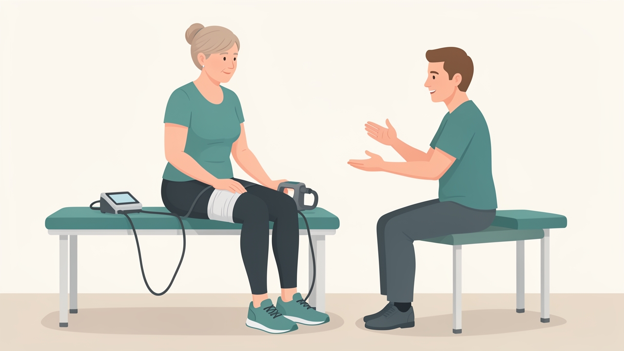
Explain the sensation beforehand: burning, fatigue and a tight cuff are expected. Sharp pain, tingling or numbness are not.
Summary
BFR therapy uses a proximal cuff to partially restrict arterial inflow and impede venous return, creating metabolic stress, cell swelling, early fast-twitch recruitment and anabolic signalling at loads of only 20–30% 1RM. Safe use depends on thorough screening, individual LOP measurement, pressures of 40–80% LOP chosen for the limb and cuff, and the standard 30-15-15-15 protocol performed two to three times weekly. Treatment is progressed through stages from post-operative activation to blended heavy loading and return to sport, guided by girth, strength LSI, hop tests and patient-reported outcomes. Contraindications such as previous VTE and vascular disease must always be respected.
References
- Patterson SD, Hughes L, Warmington S, et al. Blood flow restriction exercise: considerations of methodology, application, and safety. Front Physiol. 2019;10:533.
- Hughes L, Paton B, Rosenblatt B, Gissane C, Patterson SD. Blood flow restriction training in clinical musculoskeletal rehabilitation: a systematic review and meta-analysis. Br J Sports Med. 2017;51(13):1003–1011.
- Hughes L, Rosenblatt B, Haddad F, et al. Comparing the effectiveness of blood flow restriction and traditional heavy load resistance training in the post-surgery rehabilitation of anterior cruciate ligament reconstruction patients: a UK National Health Service randomised controlled trial. Sports Med. 2019;49(11):1787–1805.
- Slysz J, Stultz J, Burr JF. The efficacy of blood flow restricted exercise: a systematic review and meta-analysis. J Sci Med Sport. 2016;19(8):669–675.
- Takarada Y, Takazawa H, Sato Y, et al. Effects of resistance exercise combined with moderate vascular occlusion on muscular function in humans. J Appl Physiol. 2000;88(6):2097–2106.
- Loenneke JP, Wilson JM, Marín PJ, Zourdos MC, Bemben MG. Low intensity blood flow restriction training: a meta-analysis. Eur J Appl Physiol. 2012;112(5):1849–1859.
- Nakajima T, Kurano M, Iida H, et al. Use and safety of KAATSU training: results of a national survey. Int J KAATSU Training Res. 2006;2(1):5–13.
- Fujita S, Abe T, Drummond MJ, et al. Blood flow restriction during low-intensity resistance exercise increases S6K1 phosphorylation and muscle protein synthesis. J Appl Physiol. 2007;103(3):903–910.