Learning Objectives
By the end of this chapter you should be able to describe the functional anatomy of the anterior cruciate ligament (ACL), explain how its mechanics relate to injury mechanisms, recognise typical clinical presentations, select and interpret core assessments and outcome measures, and outline the principles that guide staged rehabilitation.
Anatomy and Biomechanical Function
The ACL is an intra-articular but extrasynovial structure arising from the posteromedial aspect of the lateral femoral condyle and inserting on the anterior intercondylar area of the tibia. It comprises two principal functional bundles. The anteromedial (AM) bundle is taut in flexion and is the primary restraint to anterior tibial translation at mid-to-deep flexion. The posterolateral (PL) bundle is taut in extension and contributes to rotational stability and to limiting internal rotation. The ligament is richly innervated with mechanoreceptors (Ruffini endings, Pacinian corpuscles, free nerve endings), which contribute to joint proprioception and reflexive muscle activation.
Biomechanically, the ACL is the primary restraint to anterior tibial translation and a secondary restraint to varus, valgus, and internal rotation. Its contribution changes with knee flexion angle and with the activity of the surrounding musculature. Quadriceps contraction in early flexion produces anterior tibial shear, which the ACL must resist; hamstring co-contraction reduces this load. Understanding this interplay is central to rehabilitation: the ACL is loaded not only by ligamentous strain but by muscular and neuromuscular control of the lower limb as a whole.
Injury Mechanism and Pathophysiology
Most ACL injuries are non-contact. The classic mechanism involves deceleration, a pivot or cutting manoeuvre, and a valgus-internal rotation moment at the knee, often with the foot planted and the knee near full extension to 30° of flexion. Dynamic valgus collapse, reduced hip control, and altered landing mechanics increase risk. Contact mechanisms, such as a direct blow to the lateral knee, account for a minority of cases.
Pathophysiologically, the injury produces haemarthrosis from disruption of intra-articular vasculature and a rapid inflammatory cascade. Fibres of the ligament undergo progressive failure and, unlike extra-articular ligaments, the ACL has poor intrinsic healing capacity, so surgical reconstruction is often considered for active individuals. Secondary damage to menisci and articular cartilage is common, and the bone bruising seen on MRI, particularly on the lateral femoral condyle and posterolateral tibial plateau, reflects the pivot-shift mechanism. Long-term consequences include post-traumatic osteoarthritis, with radiographic knee osteoarthritis reported in a substantial proportion of individuals at 10 to 15 years after injury (Lohmander et al., 2007).
Clinical Presentation
Patients typically describe a "pop," followed by rapid swelling (haemarthrosis) within 2 to 4 hours. Pain is often significant initially but may settle within days, leading some patients to underestimate the injury. Common complaints include giving way, a sense of instability during pivoting, reduced confidence in sport, and difficulty with stairs or descending slopes. Quadriceps weakness and arthrogenic muscle inhibition (AMI) are present early and may persist for months. Clinicians should note that effusion, pain, and AMI together limit both knee extension and quadriceps activation, which is a key early target.
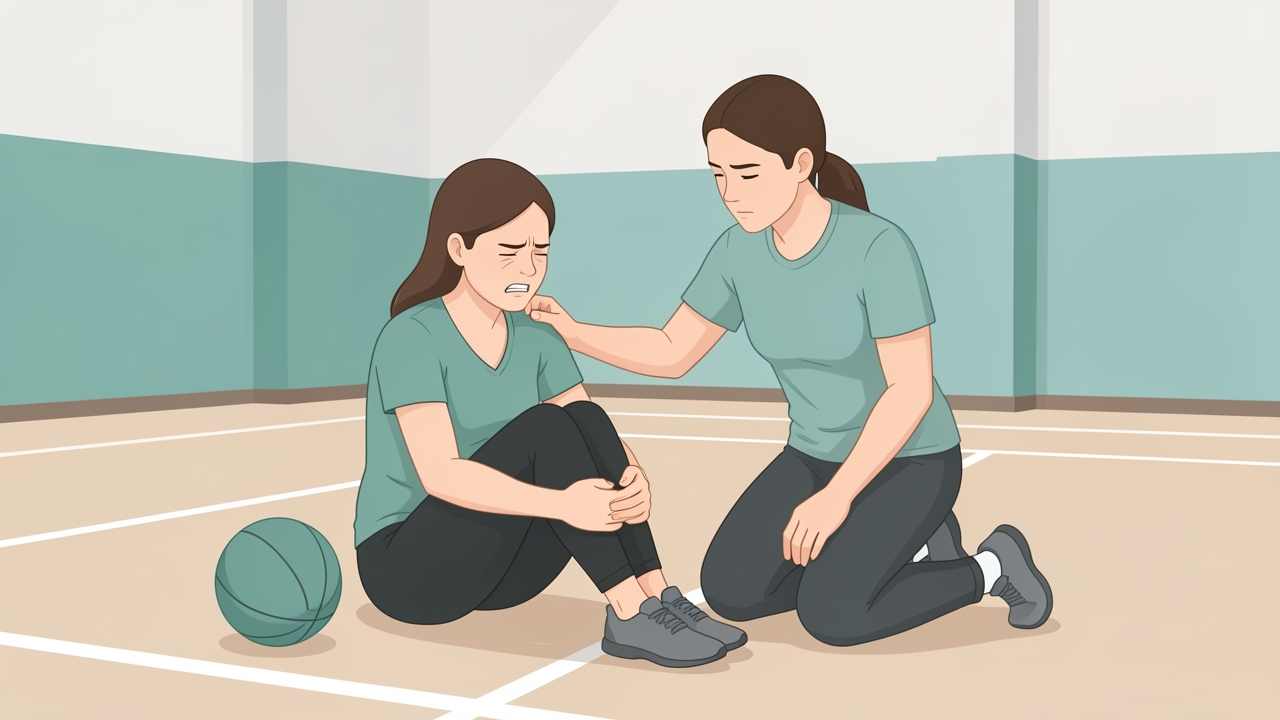
Patients typically describe a "pop", then rapid swelling within 2 to 4 hours. Pain may settle within days, so some underestimate the injury.
Clinical Assessment
A structured assessment should document the mechanism, time since injury, history of previous knee injury, and current symptoms and function.
Observation and palpation: Assess effusion (e.g., the patellar tap, or the stroke/sweep test for smaller effusions), joint line tenderness, and the presence of an extension lag. Measure girth at a fixed distance above the superior pole of the patella for quantitative tracking.
Range of motion: Measure active and passive knee extension and flexion with a goniometer. Full extension (0°) is a critical early goal; a persistent extension deficit of more than 3 to 5° is associated with poorer long-term outcome and should prompt review.
Ligament tests:
- Lachman test (30° flexion, with the tibia translated anteriorly while the femur is stabilised) is the most sensitive clinical test for ACL injury. Side-to-side difference is graded: 0 to 5 mm (grade I), 5 to 10 mm (grade II), above 10 mm (grade III), with an endpoint assessment (firm vs soft).
- Anterior drawer at 90° flexion is less sensitive and is more affected by PCL and posterior horn meniscal injury.
- Pivot shift (graded 0 to 3) assesses rotatory instability; it is often limited by pain and guarding in the acute phase and may be performed under anaesthesia.
- Instrumented laxity (KT-1000 or KT-2000 arthrometer) quantifies anterior translation. A side-to-side difference of more than 3 mm is generally considered abnormal.
Neuromuscular and strength testing: Assess quadriceps activation using a quadriceps active test or the superimposed burst technique where available. Measure isometric quadriceps and hamstring strength with a hand-held or isokinetic dynamometer at 60°/s. Hamstring-to-quadriceps (H:Q) ratios of approximately 0.6 to 0.7 are commonly cited as functional targets, though the clinical meaning of H:Q ratio is debated.
Functional and movement tests: Single-leg squat and quality of movement assessment (knee valgus, trunk control, pelvic drop) identify modifiable risk factors.
Outcome Measures and Normative Values
Selecting appropriate, validated patient-reported outcome measures (PROMs) and performance tests is essential:
- IKDC Subjective Knee Form (2000): scores 0 to 100; scores above 80 are commonly used as a satisfactory threshold.
- Knee injury and Osteoarthritis Outcome Score (KOOS): five subscales covering symptoms, pain, ADL, sport/recreation, and knee-related quality of life.
- Lysholm Knee Scoring Scale: scores above 90 indicate good function.
- Tegner Activity Scale: pre-injury level of 7 or higher indicates high-level sport participation.
- ACL-Return to Sport after Injury (ACL-RSI): assesses psychological readiness; higher scores indicate greater confidence.
- Single-leg hop tests: single hop for distance, triple hop for distance, crossover hop, and 6-m timed hop. Limb Symmetry Index (LSI) is calculated as (involved/uninvolved) × 100. An LSI of at least 90% is widely used as a return-to-sport criterion, though no single threshold is universally validated.
- Quadriceps strength LSI: targets of at least 90% are common for return to sport; a threshold of 70% is often used as an earlier milestone.
- Landing Error Scoring System (LESS): video-based assessment of landing quality.
Normative data show that many athletes, even when cleared, demonstrate persistent deficits in quadriceps strength and hop performance. Patients who demonstrate strength and hop deficits at return may be at increased risk of reinjury.
Physiotherapy Management: Principles and Staging
Rehabilitation should be criterion-based rather than purely time-based. Phases overlap, and progression depends on impairment resolution, pain, effusion control, and functional readiness. The main principles are:
- Restore full knee extension early and maintain it.
- Control effusion and pain to allow neuromuscular activation.
- Address AMI using neuromuscular electrical stimulation (NMES) and high-intensity strengthening.
- Progress load and complexity gradually, from closed to open kinetic chain, and from controlled to dynamic tasks.
- Address the whole kinetic chain, including hip and trunk strength and control.
- Use objective criteria before progression to running, cutting, and return to sport.
Phase-Based Protocol
| Phase | Timeframe (approximate) | Key Goals | Example Interventions and Dosage |
|---|---|---|---|
| Pre-operative (prehabilitation) | Before surgery | Restore ROM, quadriceps activation, reduce effusion | Quad sets, straight leg raise, NMES (quadriceps), education; 1 to 2 sessions per day |
| Acute/early post-op (phase 1) | 0 to 2 weeks | Full extension, effusion control, quad activation | Weight-bearing as tolerated, heel slides, cryotherapy, NMES 2 to 3 times daily, isometrics 3 × 10 reps, hip and ankle work |
| Early strengthening (phase 2) | 2 to 6 weeks | Full ROM, progress strength, gait normalisation | Closed-chain squat to 60 to 90°, leg press, stationary cycling, proprioceptive work, 3 sets of 8 to 12 reps |
| Intermediate (phase 3) | 6 to 12 weeks | Strength to LSI 70 to 80%, balance, controlled landing | Step-ups, lunges, hamstring curls, double- and single-leg balance, low-level plyometrics, 3 to 4 sessions per week |
| Advanced (phase 4) | 12 to 16+ weeks | Power, agility, sport-specific movement | Plyometric progression, change-of-direction drills, hop testing, neuromuscular training (e.g., FIFA 11+ components) |
| Return to sport (phase 5) | Typically 9 months minimum | Pass criteria for full participation | Sport-specific training, graded return, criteria testing |
Timeframes are indicative. Earlier phases may be extended in patients with persistent effusion or ROM deficits, and graft choice (e.g., hamstring tendon, patellar tendon, quadriceps tendon) influences graft maturation and rehabilitation pacing.
Return to sport should not be based solely on time. A return-to-sport decision should integrate strength LSI (at least 90% is often targeted), hop test LSI, validated psychological readiness (ACL-RSI), and quality of movement. Evidence suggests that combining objective strength and hop criteria with psychological readiness reduces reinjury risk. In the Delaware-Oslo cohort, patients who met simple decision rules (including quadriceps strength and hop LSI thresholds, with time from surgery) had substantially lower rates of subsequent ACL injury (Grindem et al., 2016).
Red Flags and Contraindications
Clinicians should screen for and escalate the following:
- Deep vein thrombosis: unilateral calf pain, swelling, warmth, or tenderness; refer urgently for duplex ultrasound.
- Infection: fever, increasing warmth, erythema, purulent discharge, or systemic symptoms after surgery.
- Compartment syndrome or neurovascular compromise: severe pain out of proportion, tense calf, paraesthesia, or altered pulses.
- Loss of extension or persistent effusion: may indicate arthrofibrosis, mechanical block (e.g., meniscal or loose body), or graft problems; refer for medical review.
- Acute locking or sudden giving way with swelling: consider meniscal tear or graft failure.
Relative contraindications to progressive loading include uncontrolled effusion, inability to achieve a quadriceps contraction, and pain that increases markedly with activity. Aggressive plyometric or pivoting work should be withheld until neuromuscular control, strength, and effusion criteria are met.
Evidence and Clinical Pearls
The evidence base supports several key points. ACL injury carries substantial long-term risk of osteoarthritis, which provides a rationale for rigorous rehabilitation and for addressing modifiable factors (Lohmander et al., 2007). Neuromuscular training and landing mechanics retraining reduce ACL injury risk in athletes, particularly females, and aberrant landing biomechanics are predictive of subsequent injury (Hewett et al., 2005). Systematic reviews indicate that a substantial proportion of athletes return to competitive sport after reconstruction, but return rates vary with sport, level, and psychological factors (Ardern et al., 2014). Current consensus guidelines emphasise criterion-based progression and multi-domain assessment before return to sport (Filbay & Grindem, 2019; Logerstedt et al., 2018).
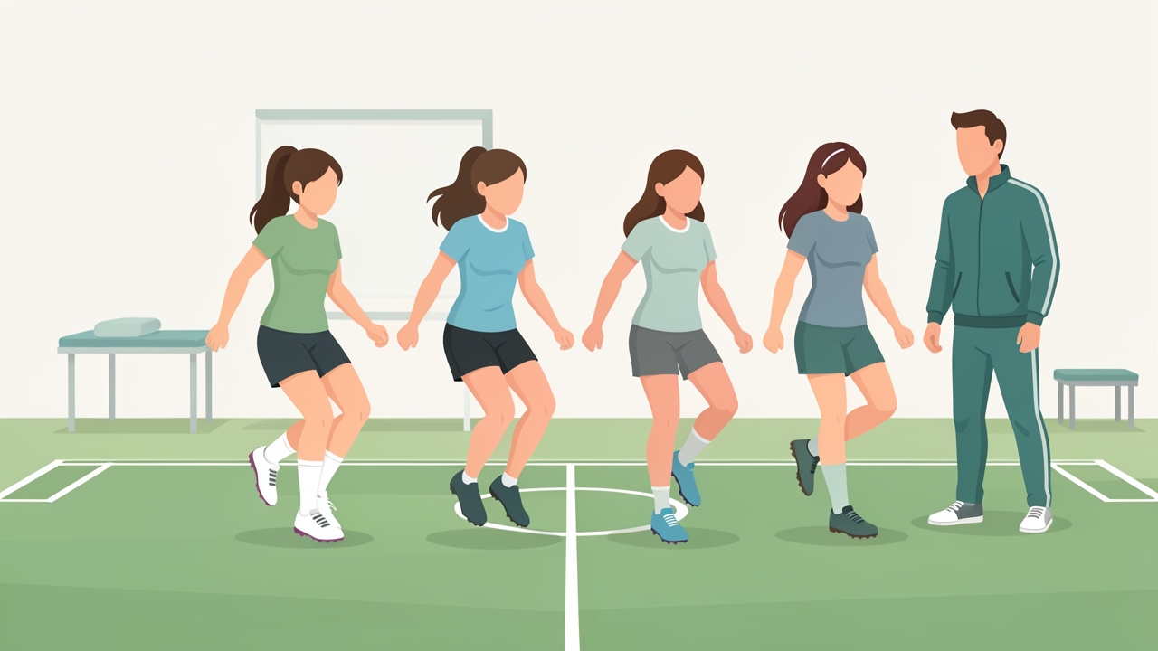
Neuromuscular training and landing mechanics retraining reduce ACL injury risk in athletes, particularly females.
Clinical pearls:
- Prioritise full extension and quadriceps activation before adding load; a knee that cannot fully extend cannot be effectively strengthened.
- Measure, document, and repeat outcomes. Trends in LSI, ROM, and effusion guide decisions more reliably than single time points.
- Do not rely on time alone for return to sport. A patient at nine months with a 70% quadriceps LSI is not ready.
- Address psychological readiness explicitly. Fear of reinjury is common and affects both performance and adherence.
- Assess the hip and trunk, not just the knee, when movement quality is abnormal.
Summary
The ACL is a key stabiliser of the knee, principally against anterior tibial translation and rotation, and its function depends on both ligamentous integrity and neuromuscular control. Most injuries are non-contact and occur during deceleration and pivoting, and they are associated with haemarthrosis, quadriceps inhibition, and risk of secondary joint damage and osteoarthritis. Accurate assessment combines clinical tests (Lachman, pivot shift, instrumented laxity), impairment measures (ROM, effusion, strength, LSI), and validated PROMs (IKDC, KOOS, Lysholm, Tegner, ACL-RSI). Rehabilitation should be staged and criterion-based, prioritising extension, effusion control, and quadriceps activation early, then progressing strength, neuromuscular control, and plyometrics before return to sport. Clinicians should maintain vigilance for red flags and apply objective, multi-domain criteria before clearing a patient to full sport participation.
References
-
Ardern, C. L., Taylor, N. F., Feller, J. A., & Webster, K. E. (2014). Fifty-five per cent return to competitive sport following anterior cruciate ligament reconstruction surgery: an updated systematic review and meta-analysis including aspects of physical functioning and contextual factors. British Journal of Sports Medicine, 48(21), 1543–1552.
-
Filbay, S. R., & Grindem, H. (2019). Evidence-based recommendations for the management of anterior cruciate ligament (ACL) rupture. Best Practice & Research Clinical Rheumatology, 33(1), 33–47.
-
Grindem, H., Snyder-Mackler, L., Moksnes, H., Engebretsen, L., & Risberg, M. A. (2016). Simple decision rules can reduce reinjury risk by 84% after ACL reconstruction: the Delaware-Oslo ACL cohort study. British Journal of Sports Medicine, 50(13), 804–808.
-
Hewett, T. E., Myer, G. D., Ford, K. R., Heidt, R. S., Colosimo, A. J., McLean, S. G., van den Bogert, A. J., Paterno, M. V., & Succop, P. (2005). Biomechanical measures of neuromuscular control and valgus loading of the knee predict anterior cruciate ligament injury risk in female athletes: a prospective study. The American Journal of Sports Medicine, 33(4), 492–501.
-
Logerstedt, D. S., Snyder-Mackler, L., Ritter, R. C., et al. (2018). Knee Stability and Movement Coordination Impairments: Knee Ligament Sprain Revision 2018. Journal of Orthopaedic & Sports Physical Therapy, 48(2), A1–A50.
-
Lohmander, L. S., Englund, P. M., Dahl, L. L., & Roos, E. M. (2007). The long-term consequence of anterior cruciate ligament and meniscus injuries: osteoarthritis. The American Journal of Sports Medicine, 35(10), 1756–1769.
-
Kyritsis, P., Bahr, R., Landreau, P., Miladi, R., & Witvrouw, E. (2016). Likelihood of ACL graft rupture: not meant to be? British Journal of Sports Medicine, 50(4), 246–251.