Clinical learning ·
Circle of Willis Explained for Physio Students
The Circle of Willis in five facts: its history, blood supply, segments, common variations and the ACom aneurysm red flag, plus five exam-style MCQs.

physioai
Instagram post
1/9
The Circle of Willis is the arterial ring at the base of the brain, the brain's backup circuit for blood flow. This article covers the five facts examiners keep coming back to, then tests you with five exam-style MCQs.
Key takeaways
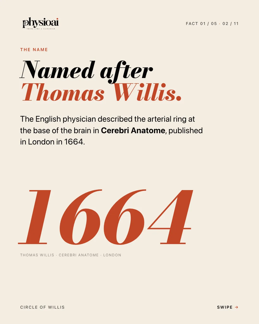
- The ring is named after Thomas Willis, who described it in Cerebri Anatome, published in London in 1664.
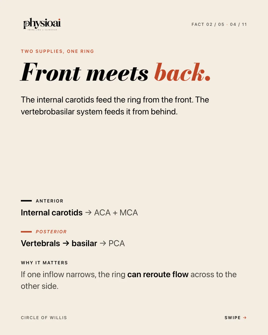
- The internal carotids feed the ring from the front and the vertebrobasilar system feeds it from behind.
- The circle is a chain of linked segments, and examiners want every link.
- A complete textbook ring is the exception, so collateral backup is not guaranteed.
- The anterior communicating artery is the most common aneurysm site in the circle.
Fact 1: The name
The Circle of Willis is named after Thomas Willis. The English physician described the arterial ring at the base of the brain in Cerebri Anatome, published in London in 1664.
Fact 2: Front meets back
The ring joins two separate blood supplies.
- Anterior circulation. The internal carotid arteries feed the ring from the front and give rise to the anterior cerebral artery (ACA) and the middle cerebral artery (MCA).
- Posterior circulation. The two vertebral arteries join to form the basilar artery, which divides into the posterior cerebral arteries (PCA).
Why it matters: if one inflow narrows, the ring can reroute blood across to the other side. This is the basis of its role as a collateral pathway.
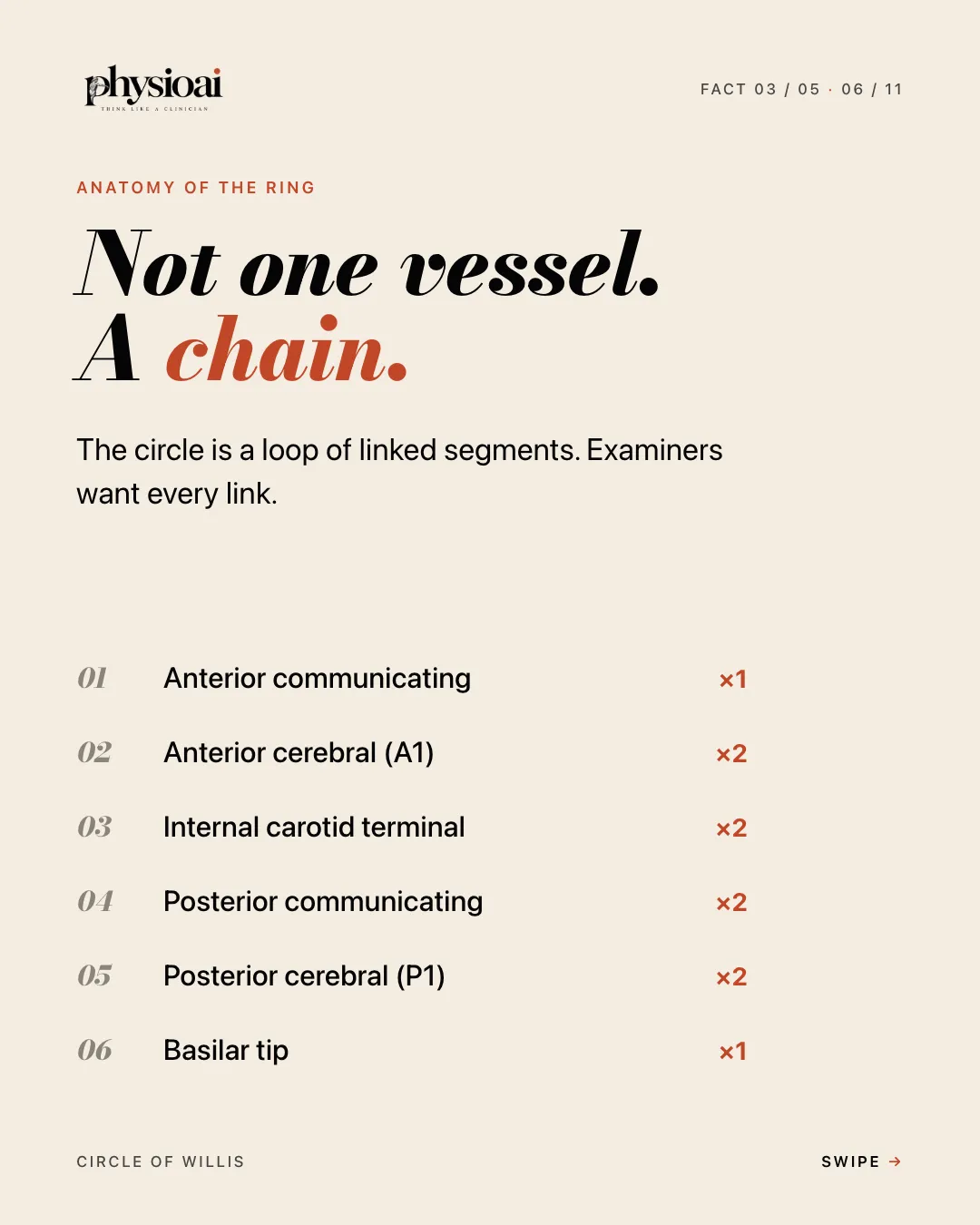
Fact 3: Not one vessel, a chain
The circle is a loop of linked segments. Learn every link and how many there are:
- Anterior communicating artery (×1)
- Anterior cerebral artery, A1 segment (×2)
- Internal carotid artery, terminal part (×2)
- Posterior communicating artery (×2)
- Posterior cerebral artery, P1 segment (×2)
- Basilar tip (×1)
A useful way to remember it is to trace the loop from front to back: the ACom joins the two ACAs, each ACA connects to its internal carotid, each internal carotid connects through a PCom to the PCA on the same side, and both PCAs meet at the tip of the basilar artery.
Note what is missing from the list. The middle cerebral artery takes blood from the ring but is not one of its segments. This is a favourite exam trap.
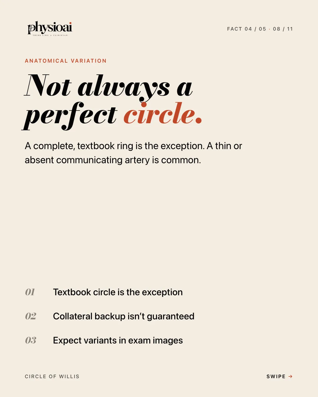
Fact 4: Not always a perfect circle
A complete, textbook ring is the exception rather than the rule. A thin or absent communicating artery is common.
- The textbook circle is the exception.
- Collateral backup is not guaranteed.
- Expect variants in exam images.
Clinically, this helps explain why two patients with a similar blockage can have very different outcomes. Whether the ring can compensate depends on the anatomy that particular person has.
Fact 5: Watch the ACom
The anterior communicating artery is the most common aneurysm site in the circle.
- Aneurysms favour branch junctions.
- A junction is a haemodynamic weak point.
- The ACom tops the circle's list.
Red flag: a sudden "worst headache of my life" should make you think of subarachnoid haemorrhage. Refer urgently. For a physiotherapist, this is a must-not-miss presentation. Stop treatment and arrange urgent medical assessment.
Why physiotherapists need this
Neuroanatomy can feel distant from day-to-day rehabilitation, but vascular territories are how you link a stroke to its signs. When you know which artery supplies which part of the brain, the pattern of weakness, sensory loss or visual loss in front of you starts to make sense. That helps you read medical notes, anticipate deficits and set realistic goals in neurological rehabilitation.
Self-check: five MCQs
Try each question before you read the answer.
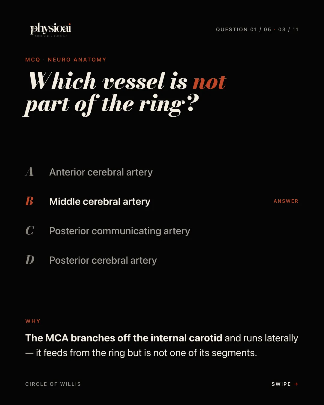
Question 1. Which vessel is not part of the ring?
- A. Anterior cerebral artery
- B. Middle cerebral artery
- C. Posterior communicating artery
- D. Posterior cerebral artery
Answer: B. Middle cerebral artery. The MCA branches off the internal carotid and runs laterally. It feeds from the ring but is not one of its segments.
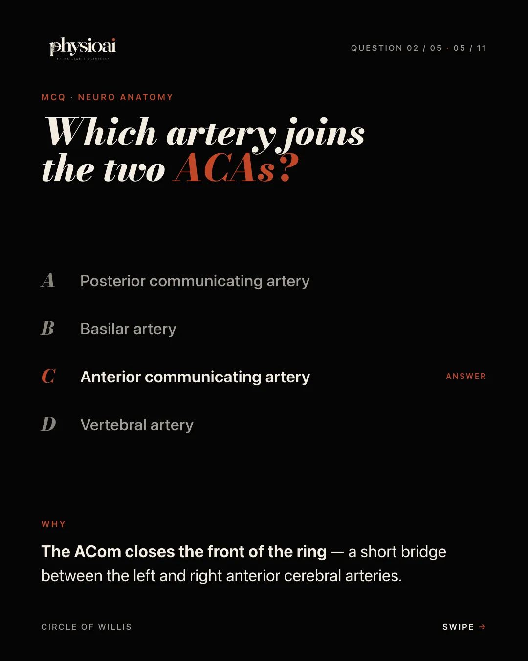
Question 2. Which artery joins the two ACAs?
- A. Posterior communicating artery
- B. Basilar artery
- C. Anterior communicating artery
- D. Vertebral artery
Answer: C. Anterior communicating artery. The ACom closes the front of the ring: a short bridge between the left and right anterior cerebral arteries.
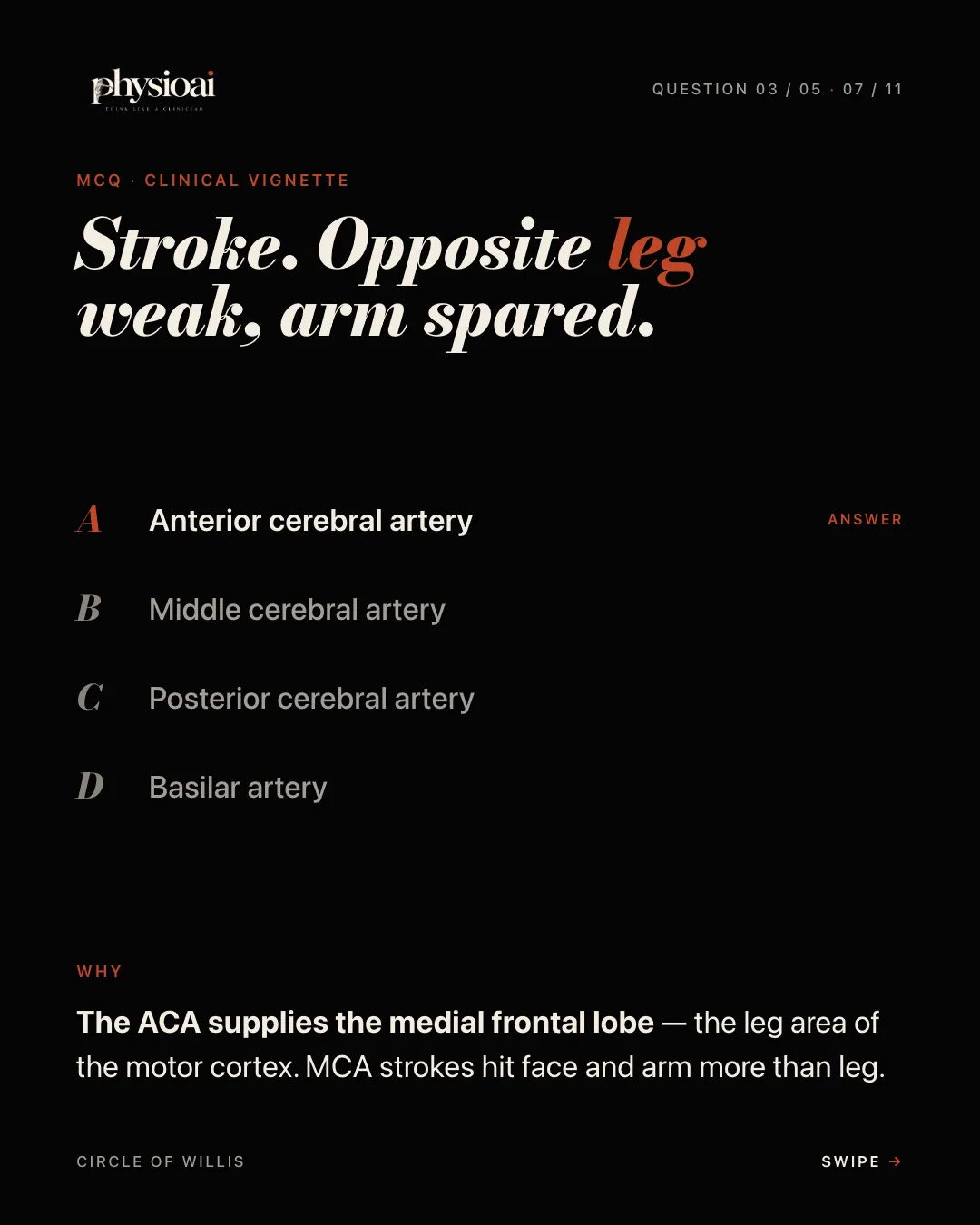
Question 3. Stroke. Opposite leg weak, arm spared. Which artery?
- A. Anterior cerebral artery
- B. Middle cerebral artery
- C. Posterior cerebral artery
- D. Basilar artery
Answer: A. Anterior cerebral artery. The ACA supplies the medial frontal lobe, the leg area of the motor cortex. MCA strokes hit face and arm more than leg.
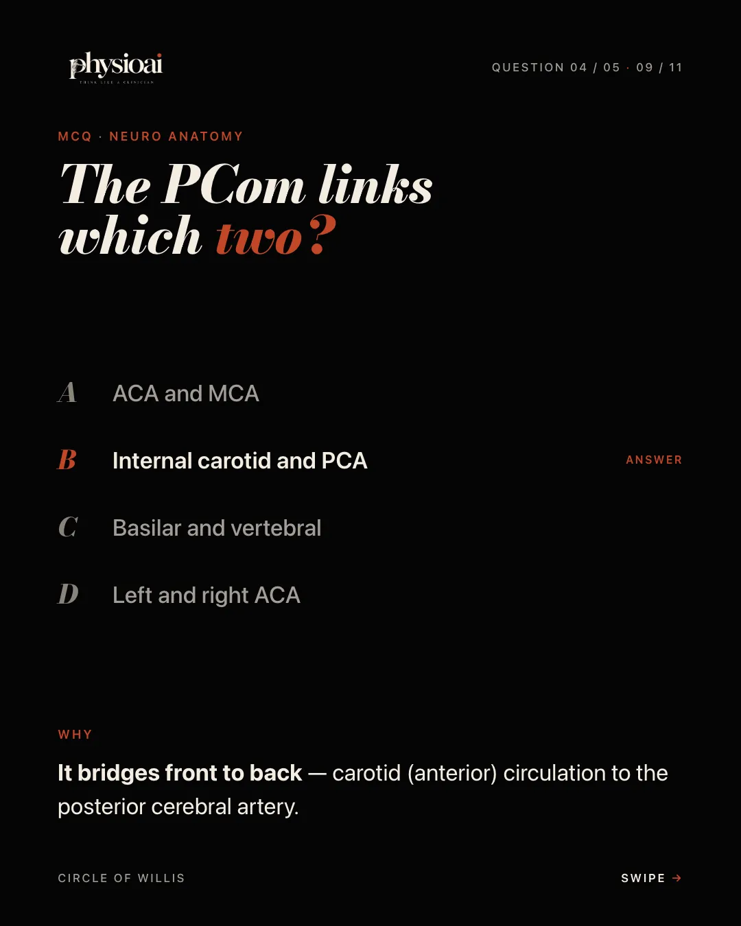
Question 4. The PCom links which two?
- A. ACA and MCA
- B. Internal carotid and PCA
- C. Basilar and vertebral
- D. Left and right ACA
Answer: B. Internal carotid and PCA. It bridges front to back, connecting the carotid (anterior) circulation to the posterior cerebral artery.
Question 5. Same half of vision lost in both eyes. Which artery?
- A. Anterior cerebral artery
- B. Middle cerebral artery
- C. Anterior communicating artery
- D. Posterior cerebral artery
Answer: D. Posterior cerebral artery. The PCA feeds the occipital lobe. Infarct leads to contralateral homonymous hemianopia, often with macular sparing.
Keep practising with PhysioAI
PhysioAI publishes fresh exam-style MCQs every day. Daily Dose is a free daily set of MCQs and flashcards that rotates across licensing exams, and Daily 45 adds 45 fresh, individually timed MCQs every morning for NPTE, CPTE or DHA. For a structured route through vascular anatomy and stroke localisation, see the Comprehensive Neuroanatomy Course in the course catalogue.